
|
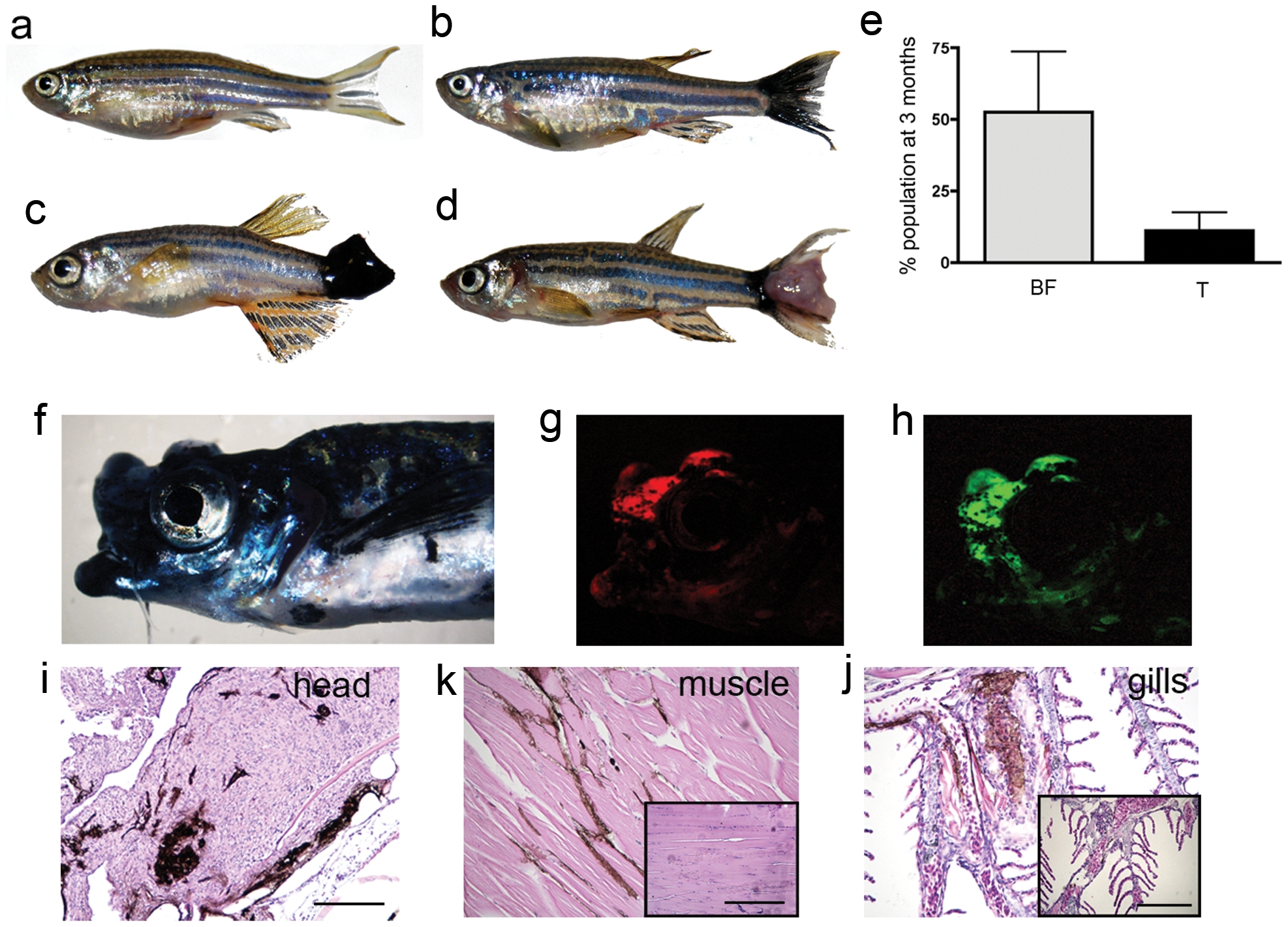
Fig. 2 kita-GFP-RAS fish develop melanoma.
a–d) Different phenotypes in three-month old kita-GFP-RAS zebrafish: a) control fish; b) black caudal fin; c) large hyper-pigmented tumor; d) hypo-pigmented tumor. e) Percentage of the progeny of kita-GFP-RAS fish incross displaying a black fin phenotype (BF) or tumor (T) at 3 months (mean±SD). f) Lateral view of a fish bearing an infiltrating melanoma of the mouth and head, which also expresses mCherry (g) and GFP-RASV12 (h). H&E staining of parraffin sections of the fish in f) that display infiltrations of melanocytes in the head (i), muscles (k), gills (j). Insets are from control animals. Calibration bars for i–j = 100 μm.