
|
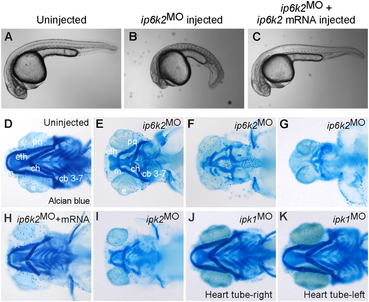
Fig. 1 IP6K2 activity is required for normal development of craniofacial and somite structures. (A–C) Lateral view of uninjected (A), ip6k2SPMO-injected (B), and ip6k2SPMO + ip6k2 mRNA-coinjected (C) live embryos at 24 hpf. Eighty percent of the ip6k2SPMO embryos had a small head and reduced trunk; both the defects were rescued by ip6k2 mRNA coinjection. Effective ip6k2SPMO suppression of splicing was confirmed by RT-PCR (Fig. S3 A and B). (D–K) Ventral view of Alcian blue stained head skeleton of 5-d-old uninjected control (D), ip6k2SPMO (E–G), ip6k2SPMO + ip6k2 mRNA (H), ipk2SPMO (I), and ipk1MO1 (J and K)-injected embryos. The anterior neurocranium was mostly maintained in 40% of ip6k2MO embryos, but distorted or reduced in 60% of embryos (F and G). The pharyngeal skeleton was affected in 92% embryos injected (n = 90). ip6k2 mRNA coinjection restored normal craniofacial skeleton (H). Craniofacial skeleton was altered in ipk2SPMO embryos (I). No craniofacial deformity was evident in ipk1MO1 embryos (J and K). e, eye; m, Meckel′s cartilage; pq, palatoquadrate; ch, ceratohyal; cb 3–7, ceratobranchial arches 3–7; eth, ethmoid plate.