
Image
|
Figure Caption
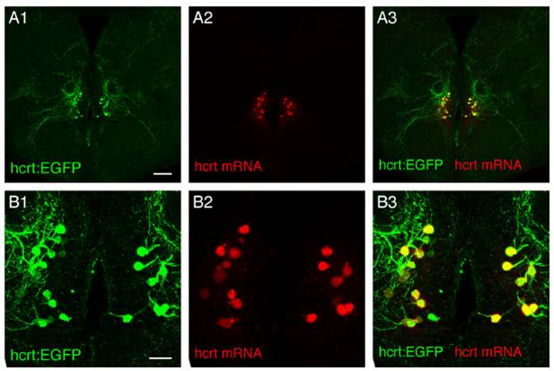
Fig. S1 (A1–A3 and associated close-ups B1–B3) Double fluorescent ISH and Immunostaining between endogenous hcrt mRNA and EGFP protein driven by hcrt promoter as visualized using confocal microscopy on brain sections of adult hcrt:EGFP transgenic fish (reconstructed stacks of 0.5- or 1-μm sections). Note the very good colocalization demonstrating the specificity of the hcrt promoter fragment used in the transgenic lines. (Scale bar, 100 μm A1–A3; 20 μm B1–B3.)
Acknowledgments
This image is the copyrighted work of the attributed author or publisher, and
ZFIN has permission only to display this image to its users.
Additional permissions should be obtained from the applicable author or publisher of the image.
Full text @ Proc. Natl. Acad. Sci. USA