
|
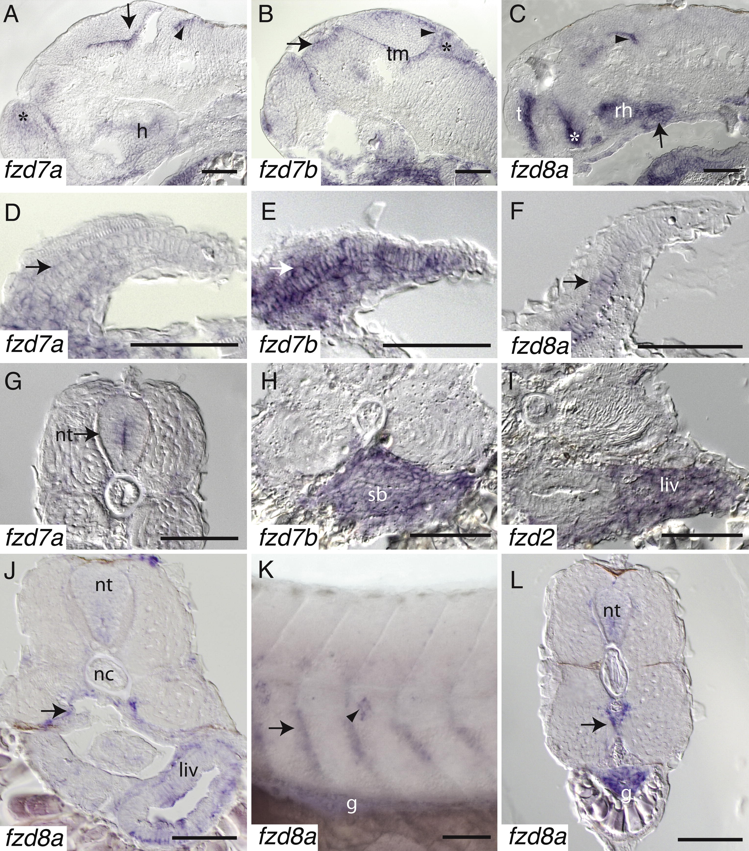
Fig. 4 Sagittal sections of 55 h post-fertilization (hpf) reveal specific expression of fzd7a, 7b, and 8a within the brain. (A) fzd7a is expressed within the dorsal hindbrain (arrowhead), forebrain (asterisk), midbrain tegmentum (arrow) and the hypothalamus. (B) fzd7b is expressed within the tectum (arrowhead), rhombic lip (asterisk), thalamus (arrow) and the tegmentum. (C) fzd8a is expressed in the telencephalon, rostal hypothalamus, along the border between the hypothalamus and the ventral thalamus (asterisk), the midbrain tegmentum (arrowhead), and the hyophysis (pituitary) (arrow). (D-F) Transverse sections of the pectoral fin bud at 55 hpf. (D) fzd7a is expressed throughout the pectoral fin bud at a low level (arrow). (E) fzd7b is expressed throughout the pectoral fin bud including the cartilage (arrow). (F) fzd8a is expressed within the cartilage of the pectoral fin bud (arrow). (G) A coronal section of a 55 hpf embryo reveals that fzd7a is expressed within the neural tube (arrow). (H–J) Coronal sections at 55 hpf (H) and 3 dpf (I–J) reveal staining within the gut. (H) fzd7b is expressed in the swim bladder and some mesenchymal tissue surrounding the gut. (I) fzd2 is expressed in the liver. (J) A more posterior coronal section demonstrates that fzd8a is expressed in the liver and along the ventral borders of the myotomes (arrow). (K) A lateral view at 3 dpf illustrates that the expression of fzd8a along the ventral borders of the myotomes is also within the anterior side of each segment of the trunk (arrow). This view also shows the expression of fzd8a within the lateral line (arrowhead) and within the gut. (L) Coronal section at 3 dpf shows expression of fzd8a in the neural tube, gut and in the vasculature (arrow). Abbreviations: (nt) neural tube, (g) gut, (nc) notochord, (liv) liver, (sb) swim bladder, (h) hypothalamus, (tm) tegmentum, (t) telencephalon, and (rh) rostal hypothalamus. Bar equals 50 μm.
Reprinted from Gene expression patterns : GEP, 9(7), Sisson, B.E., and Topczewski, J., Expression of five frizzleds during zebrafish craniofacial development, 520-527, Copyright (2009) with permission from Elsevier. Full text @ Gene Expr. Patterns