
|
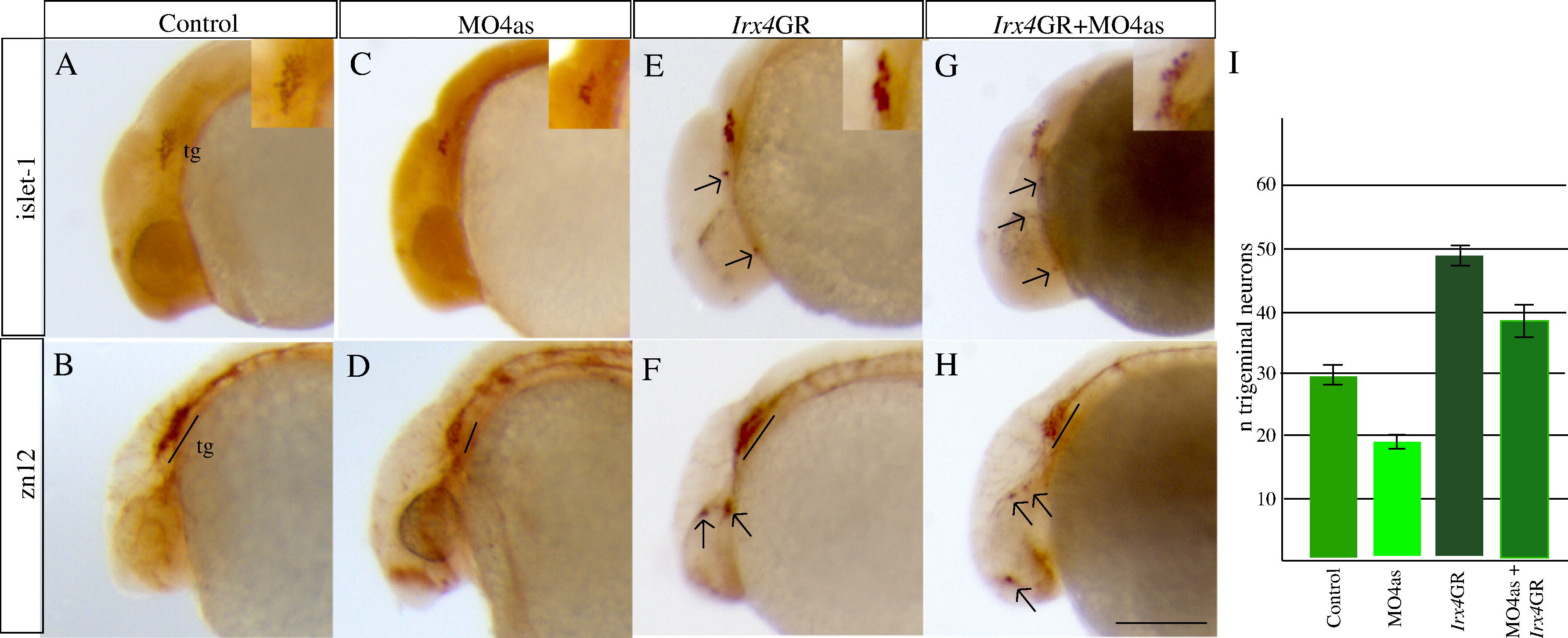
Fig. 6 irx4a regulates the number of neurons that form the trigeminal ganglion. irx4a morphant and control embryos were fixed at 24 hpf and processed for immunohistochemistry using the anti-Islet1 (A, C, E, G, insets show close-up images of the trigeminal area) or zn-12 antibodies (B, D, F, H). (A–D) Comparison of both antibody stains between control (A, B) and irx4a morphant (C, D) shows a drastic decrease in the number of neurons that form the trigeminal ganglion (compare A with C and B with D). (E, F) Gain of function experiments carried out by injection of Irx4GR mRNA show an increase in the number of trigeminal neurons (compare A with E and B with F) and occasional appearance of ectopic expression of both antibody markers in other areas of the head (arrows). (G, H) Coinjection of MO4as and Irx4GR mRNA recovers near-normal numbers of neurons in the trigeminal ganglion though there is remaining ectopic expression of Islet-1 and zn-12 antigen in the head. I, the number of anti-Islet1 labeled neurons in the trigeminal ganglia were counted in control, MO4as, Irx4GR and MO4as+Irx4GR embryos. An average for 30 embryos with S.D. was calculated. A–H show lateral views of the anterior region of embryos; dorsal is up, anterior is left. Scale bar in H: 40 μm.
Reprinted from Molecular and cellular neurosciences, 40(3), Feijoo, C.G., Saldias, M.P., De la Paz, J.F., Gómez-Skarmeta, J.L., and Allende, M.L., Formation of posterior cranial placode derivatives requires the Iroquois transcription factor irx4a, 328-337, Copyright (2009) with permission from Elsevier. Full text @ Mol. Cell Neurosci.