
|
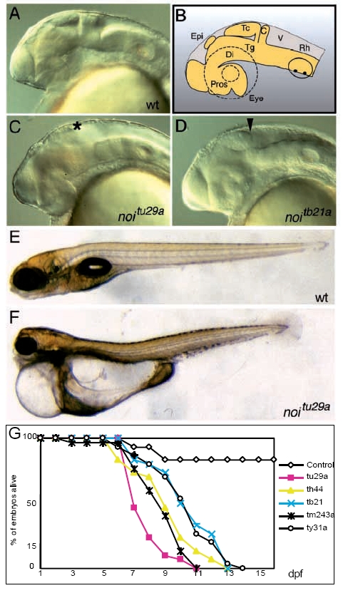
Fig. 1 Lateral views of wild-type and noi mutant embryos. (A) Wildtype embryo at 24 hours pf. (B) Schematic drawing of the structures seen in A. (C) Strong noitu29a mutant, which lacks MHB, tectum (asterisk) and cerebellum. (D) Weak noitb21 phenotype; a partially formed tectum is observed, the caudal end marked by the arrowhead. (E) A wild-type embryo at day 7. A homozygous mutant for noitu29a of the same age is seen in (F), showing severe oedema of the pericard and gut epithelium, causing delayed development. (G) Survival rates of different noi alleles. n=30 mutant embryos per allele were analyzed. The difference between the strong noitu29a allele and the weaker alleles is clearly visible. The mutants never feed and die within 2 weeks. c, cerebellum, di, diencephalon, epi, epiphysis, pros, prosencephalon, rh, rhombencephalon, tc, tectum, tg, tegmentum, v, ventricle.