
|
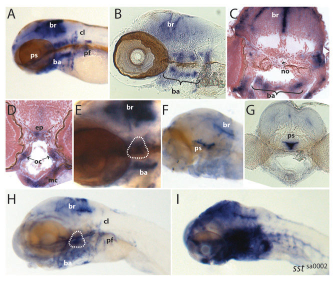
Fig. S4 Cyp26b1 expression in zebrafish. (A-E) Cyp26b1 expression in zebrafish at 3 dpf. (F-I) Cyp26b1 expression in zebrafish at 6 dpf. For sectioned material, in situ hybridization was performed on whole-mount embryos, and subsequently sections were cut using a vibratome (100 μm; B,G) or a microtome (10 μm; C,D). Expression of cyp26b1 is seen in the hindbrain, branchial arches, pectoral fins and ossified structures such as the operculum (encircled in E and H) and the parasphenoid (F,G). ba, branchial arches; br, brain; cl, cleithrum; ep, ethmoid plate; mc, meckel’s cartilage; no, notochord; oc, orbital cartilage; pf, pectoral fin; ps, parasphenoid. (F,G) Embryos that were only shortly stained show extensive staining in the parasphenoid. (H,I) Sibling (H) and sstsa0002 mutant (I) embryos showing massive upregulation of cyp26b1 expression in mutants. Identically treated sibling embryos are depicted. Genotypes were determined by sequence analysis. Other than the embryo in I, all embryos are wild type.