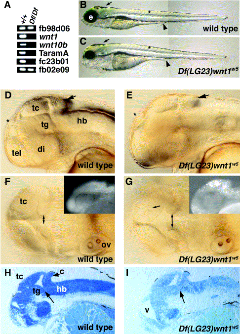
Fig. 3 Df(LG23)wnt1w5 is a deficiency for wnt1 and wnt10b. (A) PCR analysis of wild type (+/+) and homozygous mutant DNA (Df/Df). Loci indicated are from the LN54 radiation hybrid panel (see http://zfish.uoregon.edu/ZFIN for mapping panel information). (B, C) Lateral views of 5-dpf embryos, anterior to the left. Melanocytes form normally in Dfw5 embryos (arrows), somites appear normally shaped (asterisks), and the gut tube appears normal (arrowheads), indicating that tissues from all three germ layers are able to form normally. Defects appear most noticeably in the head, as Dfw5 embryos have reduced eyes (e). (D–G) Wild type and Dfw5 embryos at the prim-25 stage (36 hpf). Viewed laterally (D, E), wild type and homozygous mutants are almost indistinguishable (arrows indicate the cerebellum). When viewed dorsally (F, G), an increased distance between the medial edges of the MHB fold is visible in the mutants (double arrows). Additionally, cell death is apparent in the optic tectum (arrow in G). (Insets) Acridine orange staining of 24-hpf embryos. (H, I) Sagittal sections of 48-hpf embryos. Arrow indicates abnormal structure in the Dfw5 tegmentum– hindbrain interface. tc, tectum; tg, tegmentum; hb, hindbrain; tel, telencephalon; di, diencephalon; ov, otic vesicle; v, ventricle.
Reprinted from Developmental Biology, 254(2), Lekven, A.C., Buckles, G.R., Kostakis, N., and Moon, R.T., Wnt1 and wnt10b function redundantly at the zebrafish midbrain-hindbrain boundary, 172-187, Copyright (2003) with permission from Elsevier. Full text @ Dev. Biol.

