
|
Fig. S3
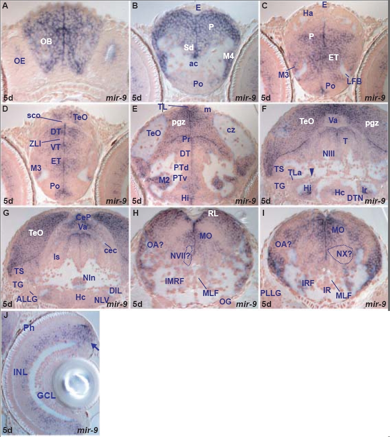
miR-9 expression in the 5dpf zebrafish brain.
miR-9 is expressed in both periventricular and adjacent cells of proliferative zones and differentiating cells arising from these domains. miR-9 expressing cells are widespread in all brain subdivisions (fore-, mid- and hind-brain) although some areas such as the larval epithalamus and the hypothalamic lateral torus are devoid of expression (Table A). In addition to the brain, miR-9 is expressed in the inner retinal nuclear layer and ciliary marginal zone. miR-9 expression is highly conserved between larval and adult brain and we find only minor differences at the regional level (Table A,F). miR-9 is expressed in a few adult habenular and hypothalamic lateral torus cells, areas devoid of larval expression.
A. transverse section through the larval rostral telencephalon at the level of the olfactory epithelium (OE) showing miR-9 expressing cells in the olfactory bulb (OB).
B. transverse section through the larval telencephalon at the level of the anterior commissure (ac) showing weakly miR-9 expressing cells in the preoptic area (Po), dorsal subpallium (Sd), migrated telencephalic area (M4) and pallium (P).
C. transverse section through the larval caudal telencephalon and rostral diencephalon showing miR-9 expressing cells in the preoptic area (Po), eminentia thalami (ET), migrated eminentia thalami (M3) and pallium (P).
D. transverse section through the larval diencephalon and rostral optic tectum showing miR-9 expressing cells in the preoptic area, (Po), eminentia thalami (ET), migrated eminentia thalami (M3), ventral thalamus (VT), dorsal thalamus (DT), zona limitans intrathalamica (ZLI) and the optic tectum (TeO).
E. transverse section through the larval diencephalon and optic tectum showing miR-9 expressing cells in the intermediate hypothalamus (Hi), ventral (PTv), dorsal (PTd) periventricular and migrated (M2) posterior tuberculum, dorsal thalamus (DT), periventricular pretectal area (Pr), proliferative zone (m), periventricular gray (pgz) zone and longitudinal torus (TL) of the optic tectum (TeO).
F. transverse section through the larval caudal hypothalamus and midbrain showing miR-9 expressing cells at the level of the lateral hypothalamic ventricular recess (lr), caudal hypothalamus (Hc), tegmental area (T, at the level of the oculomotor nucleus, NIII), semicircular torus (TS), periventricular gray zone (pgz) of the optic tectum (TeO) and cerebellar valvula (Va). The arrowhead points at the oculomotor nerve.
G. transverse section through the larval caudal hypothalamus, midbrain and isthmus showing miR-9 expressing cells in the caudal hypothalamus (Hc), diffuse nucleus of the hypothalamic inferior lobe (DIL), interpeduncular nucleus (NIn), isthmic area (Is), semicircular torus (TS), cerebellar plate (CeP), cerebellar valvula (Va) and optic tectum (TeO).
H. transverse section through the larval hindbrain at the level of the octaval ganglion (OG) showing miR-9 expressing cells in the intermediate reticular formation (IMRF), medulla oblongata (MO), presumptive octaval area (OA?) and rhombic lip (RL).
I. transverse section through the larval hindbrain at the level of the posterior lateral line ganglion (PLLG) showing miR-9 expressing cells in the inferior reticular formation (IRF), medulla oblongata (MO) and presumptive octaval area (OA?).
J. transverse section through the larval retina showing miR-9 expressing cells in the inner nuclear layer (INL) and ciliary marginal zone (arrow).