
|
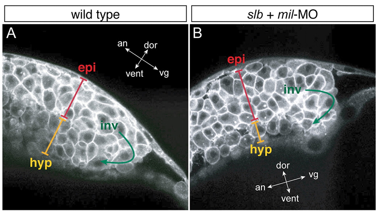
Fig. 4 slb cells with compromised Mil function undergo involution differently to WT cells. Multi-photon confocal analysis of involution cell behaviour through the shield region. Lateral views of WT embryo (A) and slb embryo injected with 4.3 ng mil-MO (B) at 60% epiboly. In the hypoblast layer of the slb embryo injected with mil-MO, cells are internalised as one- or two-cell thick layers, occasionally with space in between the cells, whereas in the WT embryo hypoblast cells are internalised as approximately three-cell thick layers throughout and are tightly packed. epi, epiblast layer; inv, involution; hyp, hypoblast layer. The orientation of embryos is shown as indicated along the animal (an)-vegetal (vg) axis and the dorsal (dor)-ventral (vent) axis.