
Image
|
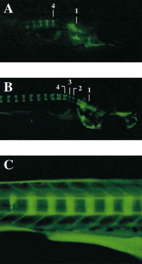
Figure Caption
Fig. 2 Visualization of calcified axial skeletal structures in developing zebrafish embryos. (A, B) Embryos at 8 and 9 dpf, respectively, showing that calcification of vertebrae number 4 appears earlier than vertebrae numbers 2 and 3. Vertebrae 1, 2, 3, and 4 are indicated. (C) Embryos at 9 dpf, showing the calcification initiation site of the vertebrae, which commences at the boundaries of each segment.
Acknowledgments
This image is the copyrighted work of the attributed author or publisher, and
ZFIN has permission only to display this image to its users.
Additional permissions should be obtained from the applicable author or publisher of the image.
Reprinted from Developmental Biology, 238(2), Du, S., Frenkel, V., Kindschi, G., and Zohar, Y., Visualizing normal and defective bone development in zebrafish embryos using the fluorescent chromophore calcein, 239-246, Copyright (2001) with permission from Elsevier. Full text @ Dev. Biol.