
|
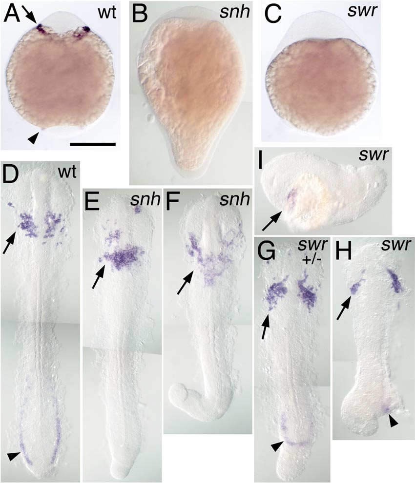
Fig. 10 Rostral expression of spi1 in BMP pathway mutants. Expression of spi1 is shown in the 8-somite stage (A–C) and 15-somite stage (D–I) embryos, in wt (A, D), snailhouse/bmp7 (snh; B, E, F), and swirl/bmp2b (swr; B, G–I) loss-of-function genetic backgrounds. Both snh and swr phenotypes are variably expressive: (E, F) Increasingly severe snh homozygous recessive phenotypes; (G) a severe swr dominant heterozygotic phenotype; (H, I) Increasingly severe swr homozygote phenotypes. (A–C) Embryos in whole mount from a ventral view, anterior to the top. (D–H) Embryos dissected from yolk and flat mounted with anterior to the top; (I) A swr embryo in lateral view, anterior to the left. Rostral expression of spi1 is marked with an arrow (A, D–I), and caudal expression is marked with an arrowhead (A, D, G, H), showing that despite the loss of the caudal domain, rostral spi1 expression is maintained in embryos exhibiting extreme “dorsalization.”
Reprinted from Developmental Biology, 246(2), Lieschke, G.J., Oates, A.C., Paw, B.H., Thompson, M.A., Hall, N.E., Ward, A.C., Ho, R.K., Zon, L.I., and Layton, J.E., Zebrafish SPI-1 (PU.1) marks a site of myeloid development independent of primitive erythropoiesis: implications for axial patterning, 274-295, Copyright (2002) with permission from Elsevier. Full text @ Dev. Biol.