
|
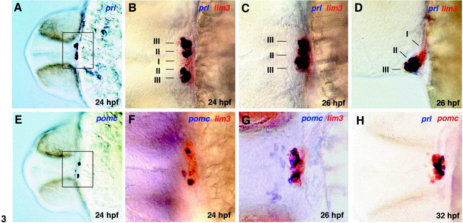
Fig. 3 Zebrafish prl and pomc are expressed in the stomodeal-hypophyseal placode. Single (A, E) and double (B–D, F–H) whole-mount in situ hybridizations with probes indicated in corresponding colors in top right corner. The age of stained embryos is indicated in bottom right corner (hpf, hours after fertilization). (D) A lateral view on the anterior region of the head; all other panels show frontal views. (B–D, F–H) are at a threefold higher magnification than (A) and (E). Frames in (A) and (E) mark magnified region. prl- and pomc-positive cells are intermingled and located in lateral (24 hpf; B, F) or anterior regions (26 hpf; D) of the lim3 domain. At 24 hpf, the number of prl-positive cells is significantly higher than the number of pomc-positive cells (compare F with B). During further development, the number of pomc-positive cells increases, while the number of prl-positive cells remains largely unchanged, so that at 32 hpf, the two cell types are present at similar numbers (H). Also, both prl- and pomc-positive cells converge from lateral regions of the pituitary anlage toward the midline, while lim3-positive cells initially located medially move posteriorly (compare B–D; see also Fig. 7 for schematic illustration; domains I, II, and III are indicated).
Reprinted from Developmental Biology, 254(1), Herzog, W., Zeng, X., Lele, Z., Sonntag, C., Ting, J.W., Chang, C.Y., and Hammerschmidt, M., Adenohypophysis formation in the zebrafish and its dependence on sonic hedgehog, 36-49, Copyright (2003) with permission from Elsevier. Full text @ Dev. Biol.