
Image
|
Figure Caption
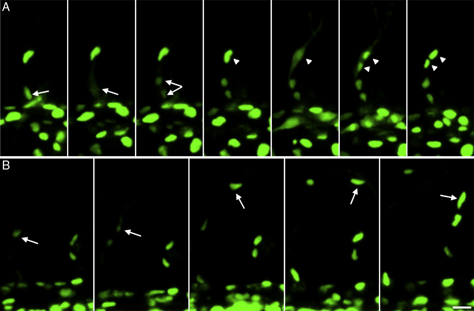
Fig. 6 Non-stereotyped behaviour of nuclei in ISVs. Picture series taken from supplementary movies 3 (A) and 4 (B) showing sprouting ISV in flk1:EGFP-NLS embryos. (A) A stalk cell (arrow) divides prior to the tip cell (arrowhead). Outline of the cell can be visualized during cell division. (B) A tip cell nucleus ends up in an adjacent ISV (arrows are following the nuclei). Scalebars: 20 μm.
Figure Data
Acknowledgments
This image is the copyrighted work of the attributed author or publisher, and
ZFIN has permission only to display this image to its users.
Additional permissions should be obtained from the applicable author or publisher of the image.
Reprinted from Developmental Biology, 316(2), Blum, Y., Belting, H.G., Ellertsdottir, E., Herwig, L., Lüders, F., and Affolter, M., Complex cell rearrangements during intersegmental vessel sprouting and vessel fusion in the zebrafish embryo, 312-322, Copyright (2008) with permission from Elsevier. Full text @ Dev. Biol.