
|
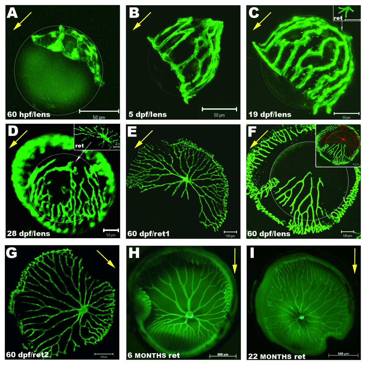
Fig. 2 Dynamic development of the hyaloid and retinal vasculature in zebrafish. Shown are fluorescent images of blood vessels on lenses and wholemount retinas dissected from Tg(fli1:EGFP) zebrafish. A: First intraocular vessels are detected at 60 hpf surrounding and attached to the forming lens. B: This vasculature evolves quickly and at 5 dpf covers the lens from the optic disk to the IOC. C: At 19 dpf an intricate network of hyaloid vessels branches around the lens. Some vessels at the posterior lens have lost contact and are attached to the retina (inset). D: At 28 dpf, detachment of the vessels from the lens has progressed anteriorly from the central region and extensive vasculature is found on the inner retina. E: Retina and F: lens from a 60 dpf zebrafish. In this specimen some vessels are attached to the lens although most of them are found on the retina. Inset in F: Overlay of the retina from E pseudo-coloured in red, and the lens from F depicts the complementing network of vessels. G: Retina of another 60 dpf zebrafish with the complete vasculature overlying the inner retina. H: Typical vascular pattern of intraocular vasculature in a 6 month old fish. I: Inner retina of a 22 month old senescent zebrafish showing slightly thinner and more fragile vessels. White circumferences demarcate the lens in A-F. Yellow arrows point from posterior to anterior lens in A-D&F and from dorsal to ventral retina in E&G-I. Scale bars: 50 μm in A-D; 100 μm in E-G and 500 μm in H-I.