
|
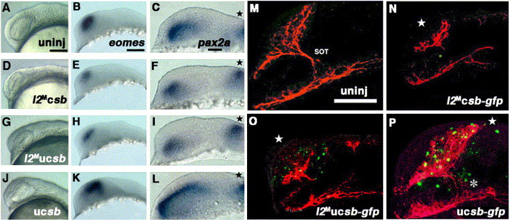
Fig. 6 Six3b cannot rescue the knockdown effect of Lhx2. (A–C) Uninjected embryos at 17 hpf. (D–F) 17-hpf embryos co-injected with lhx2-AMO plus caged-six3b mRNA that was not uncaged. (G–I) 17-hpf embryos co-injected with lhx2-AMO plus caged-six3b mRNA that was head-uncaged at 12 hpf. (J–L) 17-hpf embryos injected with caged-six3b mRNA alone that was head-uncaged at 12 hpf. (B, E, H, K) eomes expression in the dorsal telencephalon. (C, F, I, L) pax2a expression in the optic stalk. Stars point to pax2a expression at the midbrain–hindbrain boundary. (M–P) Immunostaining of the initial axonal scaffold (pseudo-colored red) and GFP fluorescence of fusion protein of Six3b and GFP (Six3b:GFP) (pseudo-colored green) at 26 hpf in the uninjected embryo (M), the embryo co-injected with lhx2-AMO plus caged-six3b:gfp mRNA that was not uncaged (N), co-injected with lhx2-AMO plus caged-six3b:gfp mRNA that was uncaged at 12 hpf (O) or injected with caged-six3b:gfp mRNA alone that was uncaged at 12 hpf (P). Stars in panels N and O show the reduction in the number of neurons in the telencephalon, and the asterisk in panel P indicates the ectopic generation of SOT in six3b:gfp-uncaged embryos as well as dorsal expansion of neurons in the telencephalon (star), showing a similar phenotype in Fig. 5P. Scale bar, 100 μm.
Reprinted from Developmental Biology, 287(2), Ando, H., Kobayashi, M., Tsubokawa, T., Uyemura, K., Furuta, T., and Okamoto, H., Lhx2 mediates the activity of Six3 in zebrafish forebrain growth, 456-468, Copyright (2005) with permission from Elsevier. Full text @ Dev. Biol.