
Fig. 3
|
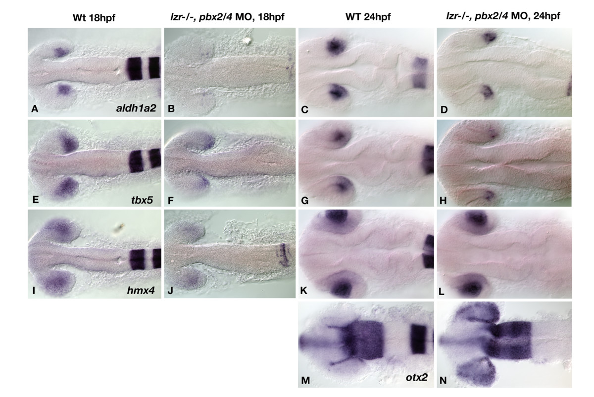
Fig. 3 Gene expression analysis via in situ hybridization shows altered expression of Pbx dependent transcripts in pbx2/4 null embryos. The expression of aldh1a2, tbx5, and hmx4 is reduced in pbx2/4 null embryos (B, F, and J), when compared to wild type (A, E, and I) at 18 hpf. At 24 hpf, aldh1a2 and tbx5 show reduced expression in pbx2/4 null embryos (D and H) when compared to wildtype (C and G), while the expression of hmx4 is unaffected (K and L). The expression of otx2 is limited to the RPE at the periphery of the eye in wild type embryos at 24 hpf (M), and is expanded to the posterior retina in pbx2/4 null embryos (N). All embryos were hybridized with the probe indicated, as well as egr2b, as an indicator of the level of Pbx function.