
|
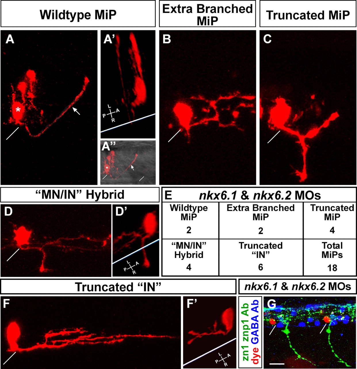
Fig. 4 MiPs become more interneuron-like in the absence of Nkx6 proteins. (A-F′) Single cells in the MiP position were identified and labeled at 24 hpf as described by Eisen et al. (Eisen et al., 1989); images were captured at 28 hpf. (A) Wild-type MiP, with its characteristic dorsal axon. The diagonal white line shows the location of the overlying segment boundary (likewise in A″,B,C,D,F,G). The asterisk shows the MiP cell body. The arrows (A,A″) indicate the MiP dorsal axon. Two other cells were also labeled during micropipette penetration of the spinal cord; these are located just dorsal of the MiP cell body. (A′) 3D rotation of the confocal image. The white line indicates the ventral aspect of the spinal cord (likewise in D′ and F′). Note that the MiP dorsal axon loops around this line, showing that it extends out of the spinal cord. (A″) The same view as A, but the fluorescent image is merged with a brightfield image to show the ventral aspect of the spinal cord and the overlying segment boundaries. (B-E) Range of phenotypes of MiPs in embryos lacking Nkx6 proteins. (B) This cell has a normal ventral axon remnant, but the dorsal axon is truncated and excessively branched. (C) This cell has abnormally retained the ventral axon and the dorsal axon is truncated. (D) This cell has abnormally retained the ventral axon; the 3D rotation in D′ shows that it extends out of the spinal cord. The cell has not developed a dorsal axon, but instead has an interneuron-like axon within the spinal cord, as shown in the rotation in D′. (E) Quantification of phenotypes shown in B-D and F. IN, interneuron; MN, motoneuron. (F,F′) This cell has neither a ventral nor a dorsal axon, but has an excessively branched interneuron-like axon that is truncated relative to those of wild-type interneurons. The 3D rotation in F′ shows that this interneuron-like axon is entirely within the spinal cord. (G) Triple label showing that dye-labeled MiPs (red) are located in the normal MiP position, are adjacent to GABA-positive interneurons (blue) in the VeLD, KA′ and KA″ positions (white asterisks above cells in the VeLD position), and are in segments with normal CaP axons but lacking MiP axons as revealed by labeling with zn1 and znp1 Abs (green). Scale bar: 10 μm in A-D′,F,F&prime& 20 μm in G.