
|
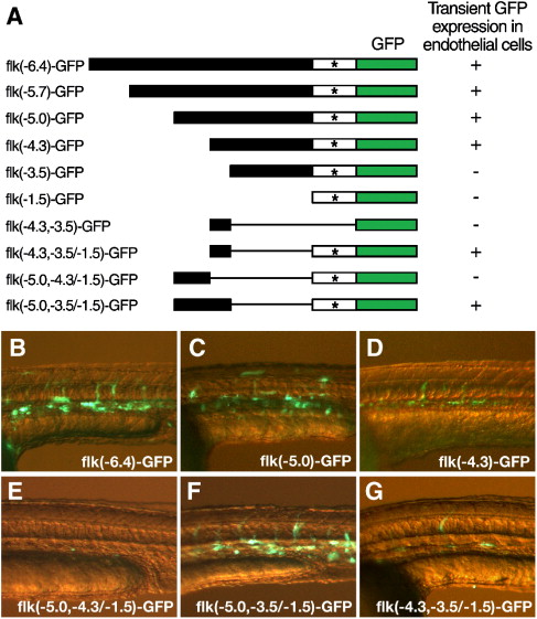
Fig. 2 Deletion analysis of the flk1 regulatory region identifies a highly conserved element that is necessary for endothelial expression. (A) Schematic diagram of the deletion constructs of flk1-GFP reporter. Linearized DNA of each construct was injected in wild type zebrafish embryos at the 1-cell stage. GFP expression in injected embryos was analyzed after 1 day of development. The transient endothelial expression directed by each construct is summarized by a plus (endothelial expression) or a minus (no detectable endothelial expression) to the right of the line representing each construct. * marks the transcription initiation site of flk1. (B–G) Transient GFP expression in endothelial cells of 1-day-old embryos injected with flk(- 6.4)-GFP (B), flk(- 5.0)-GFP (C), flk(- 4.3)-GFP (D), flk(- 5.0, - 4.3/- 1.5)-GFP (E), flk(- 5.0, - 3.5/- 1.5)-GFP (F) or flk(- 4.3, - 3.5/- 1.5)-GFP (G).
Reprinted from Developmental Biology, 304(2), Choi, J., Dong, L., Ahn, J., Dao, D., Hammerschmidt, M., and Chen, J.N., FoxH1 negatively modulates flk1 gene expression and vascular formation in zebrafish, 735-744, Copyright (2007) with permission from Elsevier. Full text @ Dev. Biol.