
|
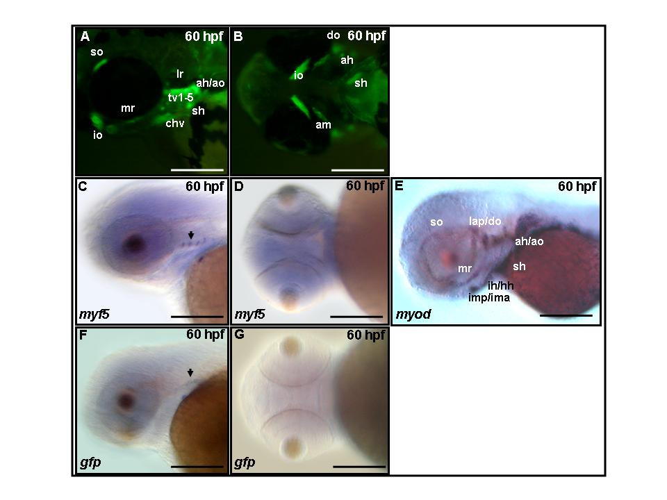
Fig. 5 Green fluorescent protein (GFP) persists in cranial muscles of Tg(myf5(80k):GFP) transgenics. (A,B) GFP fluorescence is apparent in adductor hyomandibulae (ah), adductor mandibulae (am), adductor operculi (ao), constrictor hyoideus ventralis (chv), dilatator operculi (do), inferior oblique (io), lateral rectus (lr), medial rectus (mr), sternohyoideus (sh), superior oblique (so), and transverse ventralis (tv1–5) at 60 hours postfertilization (hpf). (C, D) Endogenous myf5 transcripts and (F, G) GFP mRNA are restricted to four spots by 60 hpf using whole-mount mRNA in situ hybridization. (E) At 60 hpf, myod transcripts are detected in most if not all cranial muscles. (A,C,E,F) Side views, rostral to the left, dorsal to the top; (B,D,G) ventral view, rostral to the left. Scale bars: 200 μm in all panels.