
|
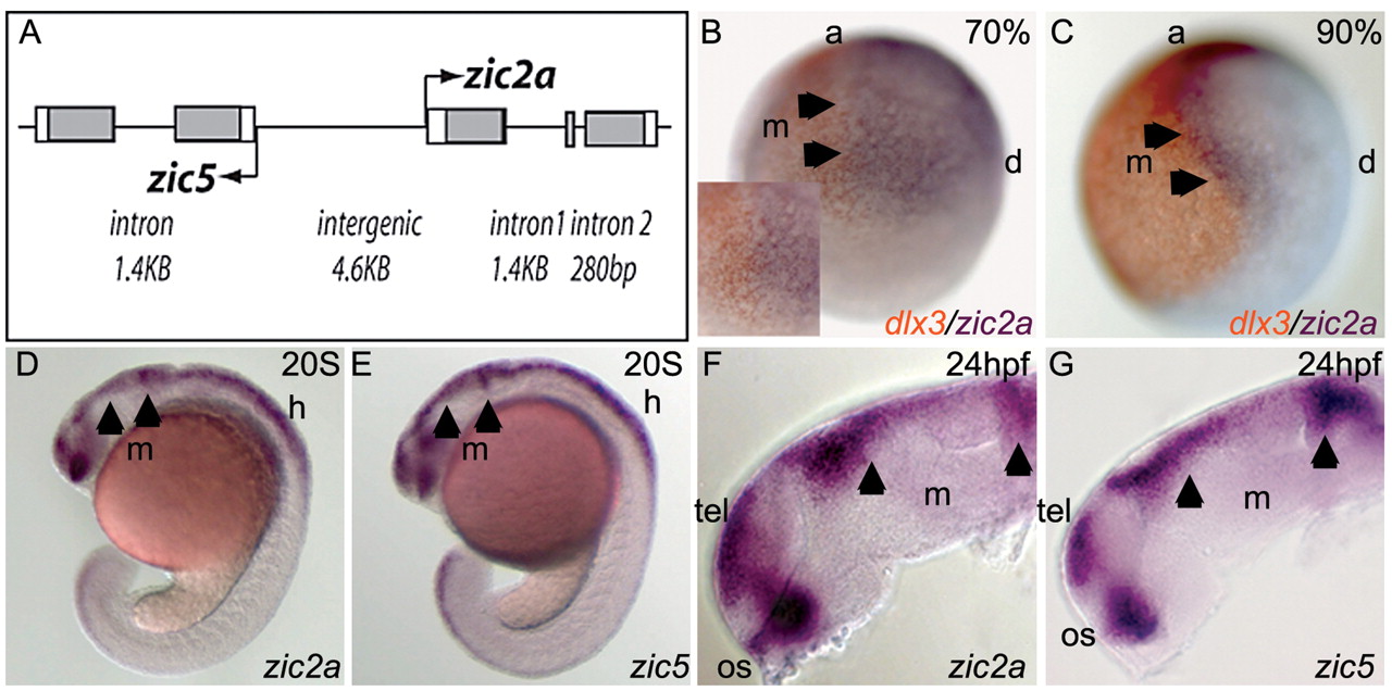
Fig. 1 Zic2a and zic5 are linked and co-expressed in presumptive dorsal brain. (A) Genomic arrangement of zic2a and zic5. Coding regions are shown as gray boxes, non-coding transcribed regions as white boxes, and introns as lines. (B-G) Embryos stained by ISH for zic2a or zic5 (purple), and dlx3 (orange). (B) At midgastrula stages, zic2a is expressed throughout neurectoderm, whereas dlx3 marks the adjacent non-neural ectoderm. (C) By late gastrula stages, zic2a is restricted to the lateral neural plate and forms a sharp border with dlx3. (D,E) During somitogenesis, zic2a and zic5 are similarly expressed in the dorsal neural tube, ventral diencephalon and optic stalks. (F,G) At 24 hpf, zic2a and zic5 are coexpressed in the optic stalk, telencephalon and dorsal diencephalon, and weakly in the tectum. All views are lateral, anterior to the left, except in B,C where anterior (a) is at the top. Arrowheads mark anterior and posterior borders of presumptive midbrain. d, dorsal; m, midbrain; h, hindbrain; os, optic stalk; tel, telencephalon.