
|
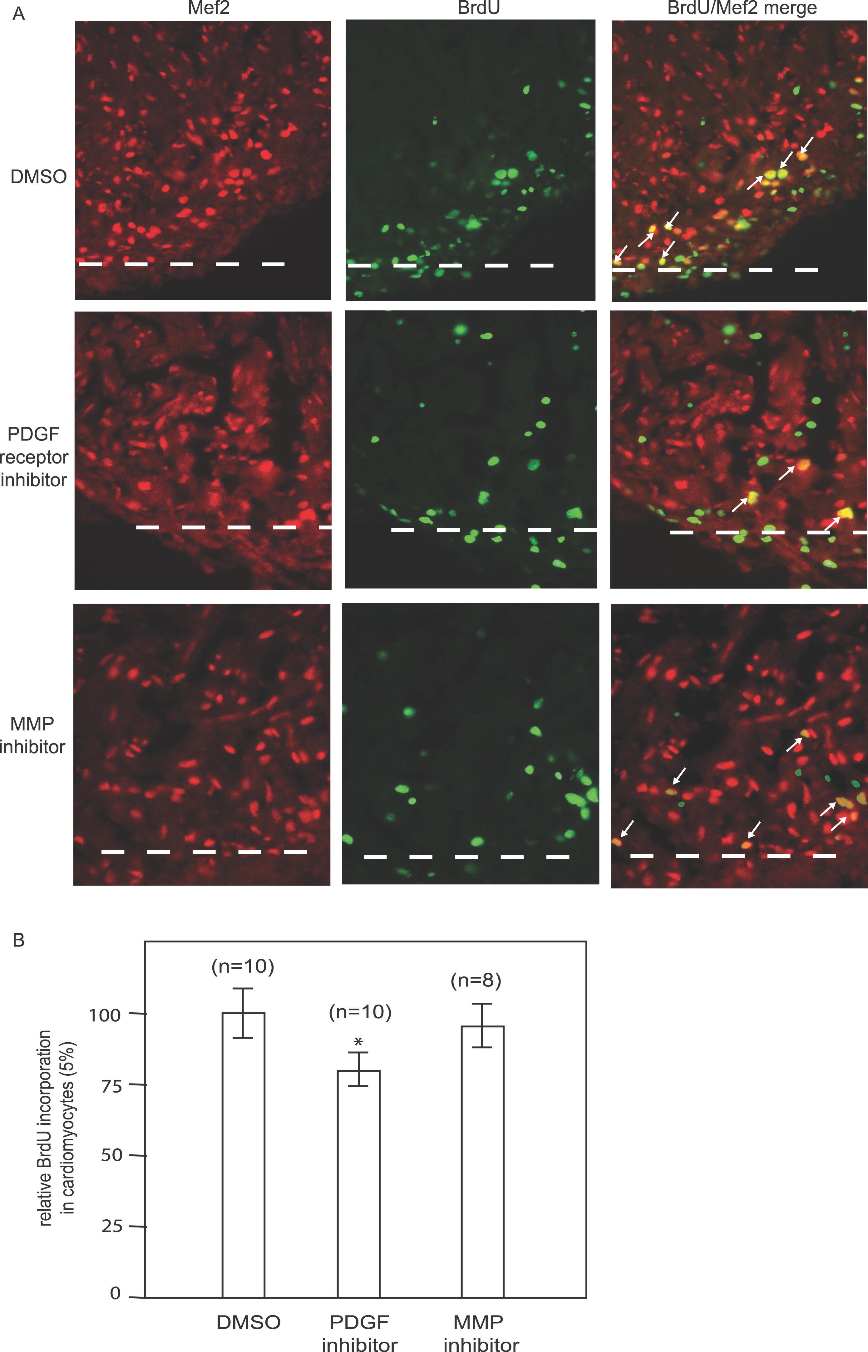
Fig. 6 PDGF Signaling Is Required for Cardiomyocyte Proliferation during Zebrafish Heart Regeneration. After heart surgery, the fish were allowed to recover for one day and were then treated with either the PDGF receptor inhibitor AG1295, DMSO as a negative control, or the MMP inhibitor GM6001 as a specificity control. (A) Heart sections were stained with MEF2 antibody (red) to visualize the cardiomyocyte nuclei and with BrdU antibody (green) to measure DNA synthesis. The BrdU/MEF2 double-positive nuclei (white arrows) are shown in yellow. Many BrdU-positive cells are not cardiomyocytes. PDGF receptor inhibitor treatment resulted in fewer BrdU-positive cardiomyocytes while DMSO and MMP inhibitor treatment did not. The dashed white line marks the amputation plane. (B) Quantification of DNA synthesis in cardiomyocytes during zebrafish heart regeneration. Results were normalized to DMSO-treated control hearts (100%). AG1295 treated hearts had 16% (p < 0.05) fewer BrdU-positive cardiomyocytes compared to DMSO control hearts (Student's t-test), whereas the difference between GM6001-treated and DMSO-treated hearts was statistically insignificant (p > 0.05). Error bars indicate standard error of the mean.