
Image
|
Figure Caption
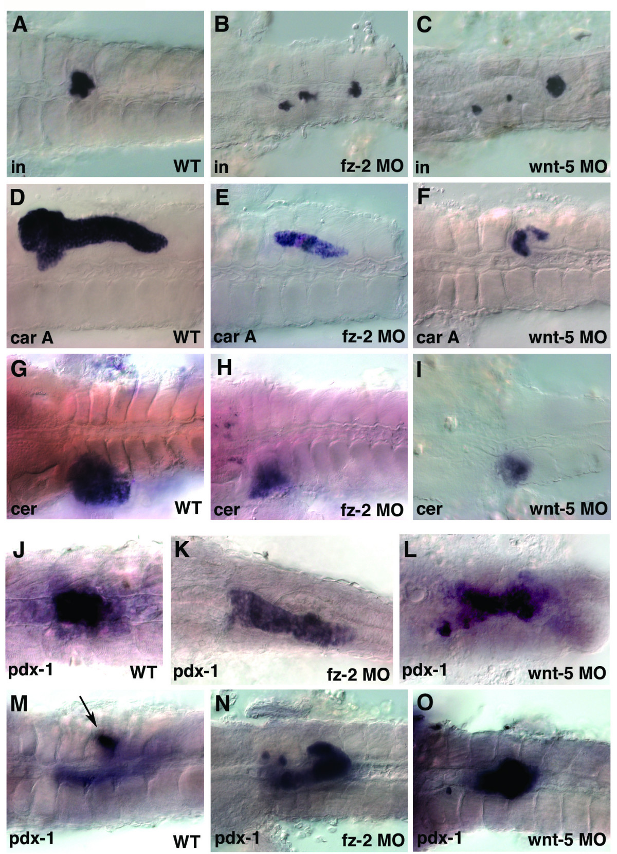
Fig. 6 Wnt-5 and Fz-2 morphant embryos have other similar defects. In all panels, view is dorsal, anterior is to the left. (A-I, M-O) 3dpf, (J-L) 24 hpf stage. (A, D, G, J, M) Wild-type embryos. (B, E, H, K, N) Fz-2 morphants. (C, F, I, L, O) Wnt-5 morphants. In situ hybridization analysis of (A-C) insulin, (D-F) carboxypeptidase A, notice the hollow spot indicating the position of the islet, (G-I) ceruloplasmin, (J-O) pdx-1, (M) arrow, pdx-1-staining in islet.
Figure Data
Acknowledgments
This image is the copyrighted work of the attributed author or publisher, and
ZFIN has permission only to display this image to its users.
Additional permissions should be obtained from the applicable author or publisher of the image.
Full text @ BMC Biol.