
|
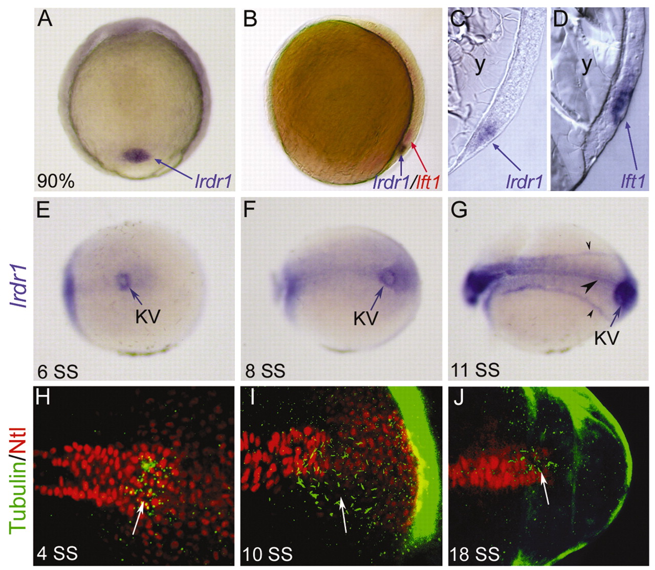
Fig. 1 lrdr1 is expressed in dorsal forerunner cells (DFCs) and in ciliated Kupffer′s vesicle (KV). (A) lrdr1 is expressed exclusively in DFCs (arrow) at 90% epiboly. Dorsal view, anterior towards the top. (B) Lateral view of an embryo (dorsal on the right) at 80% epiboly stained for lrdr1 (purple) and lft1 (red) expression. lrdr1 is restricted to DFCs, whereas lft1 is expressed in margin cells and midline cells but not DFCs. Sectioned embryos confirmed that lrdr1 (C) is expressed in only DFCs and that lft1 (D) is expressed in cells adjacent to DFCs at 90% epiboly. No expression of lrdr1 or lft1 was detected in the yolk (y). (E-G) lrdr1 RNA expression in KV (arrow) at (E) 6 SS, (F) 8 SS and (G) 11 SS. lrdr1 was also detected in the tailbud at 8 SS (F) and in the floor plate, notochord and intermediate mesoderm (arrowheads) by 11 SS (G). Posterior-dorsal views with the anterior to the left. (H-J) Immunofluorescence at (H) 4 SS, (I) 10 SS and (J) 18 SS using anti-acetylated Tubulin antibodies (green) to detect cilia and anti-ntl antibodies (red) to label nuclei in the notochord and KV. Arrows indicate cilia in KV. Embryos were transversely bisected for visualization of the tailbud.