FIGURE

Figure 3—figure supplement 2.

ID
ZDB-FIG-191230-1597
Publication
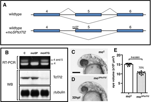
Young et al., 2019 - Developmentally regulated Tcf7l2 splice variants mediate transcriptional repressor functions during eye formation
Other Figures
All Figure Page
Back to All Figure Page
Figure 3—figure supplement 2.

Splicing specific morpholino knockdown of <italic>tcf7l2</italic> splice variants that include exon5.

(A) Cartoon showing the rationale of using moSPtcf7l2 that targets intron 4/exon five splice site boundary blocking the splicing machinery and making it skip to the next splicing acceptor site in exon 6. (B) RT-PCR (top panel) and western blot (WB, middle and bottom panels) performed using RNA and proteins extracted from 24hpf zebrafish embryos injected with: control morpholino (first lane), moSPtcf7l2 (moSP, second lane), and moATGtcf7l2 (moATG, third lane). cDNA was amplified using set ‘a’ primers (Figure 1A, 5’F1 vs Splice R1, Materials and methods). Anti human Tcf7l2 and anti-gamma tubulin antibodies were used in Western blot experiments (middle and bottom panel). (C, D) 32hpf embryos injected with 1.25 pmol of (C) control morpholino (moC) or (D) tcf7l2 exon five splicing morpholino (moSPtcf7l2). Scalebar in (C) is 200 µm. (E) Plot showing estimated eye volume of 32hpf live embryos injected with injected with moC or moSPtcf7l2 as in (C, D). Error bars are mean ± SD, P values from unpaired t test. Percentage indicates average eye profile size relative to control.

Expression Data

Expression Detail
Antibody Labeling
Phenotype Data
Fish:
Knockdown Reagent:
Observed In:
Stage: Prim-15

Phenotype Detail
Acknowledgments
This image is the copyrighted work of the attributed author or publisher, and ZFIN has permission only to display this image to its users. Additional permissions should be obtained from the applicable author or publisher of the image. Full text @ Elife