
Fig. S2
- ID
- ZDB-FIG-190919-9
- Publication
- Vogrin et al., 2019 - Evolutionary Differences in the Vegf/Vegfr Code Reveal Organotypic Roles for the Endothelial Cell Receptor Kdr in Developmental Lymphangiogenesis
- Other Figures
- All Figure Page
- Back to All Figure Page
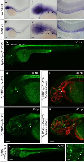
In situ hybridisation for kdr and flt4, and analysis of vegfd expression, Related to Figure 4. (A-F) In situ hybridisation using probes designed to kdr (A-C) and flt4 (D-F) showing expression in the craniofacial region as well as in the posterior cardinal vein. (G-M) Expression of vegfd visualised using the TgBAC(vegfd:eYFP)uq42bh transgenic line. (G-I) At 36 hpf, expression is observed in the craniofacial region close to where lymphatic sprouts emerge from the primary head sinus (arrows). (J,K) At 60 hpf, vegfd expression is observed in the region close to where the otolithic lymphatic sprout emerges and in the region where mural LECs sprout from the choroidal vascular plexus (arrows). (L,M) At 72 hpf continued expression of vegfd can be observed in the craniofacial region where the first lymphatic sprouts form and in branchial arches where lymphatics sprouts emerge (arrows). Scale bars in A, D and L represent 200 m, in B, C and E-K 100 m and in M 50 m. |
| Genes: | |
|---|---|
| Fish: | |
| Anatomical Terms: | |
| Stage: | Prim-25 |