
Fig. 4
- ID
- ZDB-FIG-190919-28
- Publication
- Shainer et al., 2019 - Agouti-Related Protein 2 Is a New Player in the Teleost Stress Response System
- Other Figures
- All Figure Page
- Back to All Figure Page
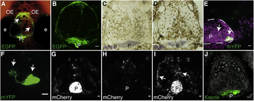
Pineal AgRP2-Expressing RPE-like Cells Are Secretory Cells (A) Dorsal view of a 21-dpf agrp2:EGFP fish head. The EGFP is not confined to the pineal and can be observed above the telencephalon (arrowheads). (B) Dissected skull of adult agrp2:EGPF fish, immunostained with an antibody against EGFP. Ventral view, anterior to top, is shown. The EGFP remains bound to the skull. Scale bar, 100 μm. (C) ISH of agrp2 (purple) performed on an adult skull. Ventral view (same as B), anterior to top, is shown. agrp2 mRNA is localized to the pineal gland. (D) ISH of egfp (purple) performed on an adult skull. Ventral view (same as B), anterior to top, is shown. egfp mRNA is localized to the pineal gland. (E) Dorsal view of a 21-dpf agrp2:Gal4-VP16; UAS:nfsB-mCherry; UAS:mYFP fish, focusing on the pineal gland with sparse mYFP labeling. The membrane-bound YFP (green) is confined to the pineal (arrowheads), and the mCherry can be found outside of the pineal, possibly secreted from the pineal region. Dashed line represents the circumference of the pineal gland. Scale bar, 10 μm. (F) Enlargement of (E) reveals the cellular architecture of AgRP2 cells, bearing branched microvilli-like structure (left arrowhead). Scale bar, 10 μm. (G) Dorsal view of a 7-dpf agrp2:Gal4-VP16; UAS:nfsB-mCherry fish larvae before laser ablation, focusing on the pineal gland and the telencephalic area. The mCherry is mainly detected in the pineal gland. Scale bar, 10 μm. (H) Same larva as (G), at 10-dpf, 3-day post laser-ablation. Dorsal view focuses on the pineal gland and the telencephalic area. The mCherry signal from the pineal is lost and is merely detected above the telencephalic region. Scale bar, 10 μm. (I) Dorsal view focusing on the pineal gland and the telencephalic area of 10-dpf agrp2:Gal4-VP16; UAS:nfsB-mCherry control larva with intact pineal gland. At this age, the mCherry signal can already be detected above the telencephalic area (arrowheads). Scale bar, 10 μm. (J) Dissected skull of adult agrp2:Gal4-Vp16; UAS:Kaede fish. Ventral view, anterior to top, is shown. The Kaede signal can only be detected in the pineal area. Scale bar, 100 μm. e, eye; OE, olfactory epithelium; P, pineal. |
| Genes: | |
|---|---|
| Fish: | |
| Condition: | |
| Anatomical Terms: | |
| Stage Range: | Days 7-13 to Adult |
| Fish: | |
|---|---|
| Condition: | |
| Observed In: | |
| Stage: | Days 7-13 |