
Fig. 1
- ID
- ZDB-FIG-190708-48
- Publication
- Kujawski et al., 2019 - penner/lgl2 is required for the integrity of the photoreceptor layer in the zebrafish retina
- Other Figures
- All Figure Page
- Back to All Figure Page
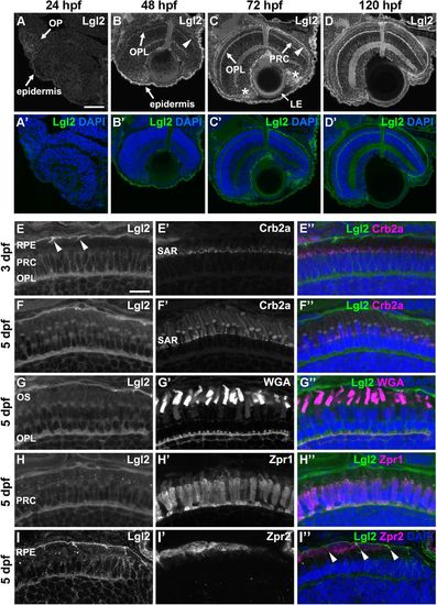
Lgl2 expression is upregulated in the retina by 72 dpf and localizes basolaterally in photoreceptor and RPE cells.Immunostaining of transverse retinal sections. (A–D′) Lgl2 expression during retinal development: A,A′, 24 hpf; B,B′, 48 hpf; C,C′, 72 hpf; D,D′, 120 hpf. Lgl2 expression is upregulated by 72 hpf in the RPE (C, arrowhead) and the OPL. Asterisks in C denote expression in the ciliary marginal zone (CMZ). OP, olfactory placode; OPL, outer plexiform layer; PRC, photoreceptor cell layer; LE, lens epithelium. Scale bar: 50 µm. (E–E″) At 3 dpf, Lgl2 (E) localizes basolaterally in the PRC and in the RPE. Lgl2 staining does not overlap with that of Crb2a (E′), which localizes to the subapical region (SAR). Arrowheads in E denote lateral Lgl2 localization in RPE cells. E″ shows merged image. (F–I″) Lgl2 localization at 5 dpf. (F–F″) Co-staining of Lgl2 with Crb2a shows that Lgl2 staining remains basolateral as PRCs mature. (G–H″) Co-staining of Lgl2 (G,H) with WGA (G′) or Zpr1 (H′) illustrates Lgl2 localization in the OPL. (I–I″) Co-staining of Lgl2 (I) with Zpr2 (I′) reveals basolateral expression in the RPE (arrowheads in I″ denote lateral localization). OS, outer segments. Scale bar: 10 µm. |