
Fig. 2
- ID
- ZDB-FIG-190404-3
- Publication
- Ton et al., 2018 - Collagen COL22A1 maintains vascular stability and mutations in COL22A1 are potentially associated with intracranial aneurysms
- Other Figures
- All Figure Page
- Back to All Figure Page
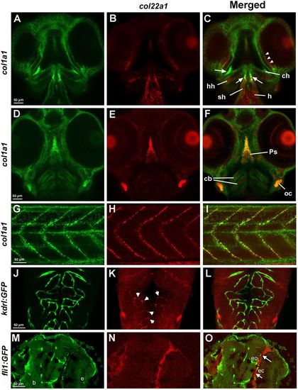
col22a1 expression in the head and trunk region analyzed by FISH and FISH/immunofluorescence. (A-I) Expression of col22a1 (red) and col1a1 (green) at 3 dpf, which commonly marks connective tissue. Expression was analyzed by HCR. In the transverse confocal sections of the cranial tissue (ventral view, A-F), col22a1 expression is apparent in the periocular tissue (arrowheads, C), craniofacial muscle, including sternohyoideus (sh) and hyohyoideus (hh) muscles, parasphenoid bone (Ps), the heart (h), presumptive chondrocytes within ceratobranchial (cb) and ceratohyal arches (ch), and connective tissue within the developing ear (otic capsule, oc). Note that the expression of both markers overlaps within the parasphenoid bone, otic tissue, connective tissue around the eyes (arrowheads, C), and the tendon/ligament attachment sites of the sternohyoideus and hyohyoideus muscles (arrows, C). Within the myotendinous junction in the trunk region (G-I), all col22a1-expressing cells were also positive for col1a1 expression. Note that col1a1 also labels keratinocytes within the epidermis. (J-L) col22a1 expression analyzed by HCR partially overlaps with vascular endothelial expression of kdrl:GFP. Cranial vasculature of whole-mounted embryos at 3 dpf was imaged dorsally by confocal microscopy. Anterior is to the top. Maximal-intensity projection of selected slices is shown. (M-O) Longitudinal cranial sections of embryos stained by WISH/immunofluorescence for col22a1 (red) and vascular endothelial fli1:GFP (green) at 72 hpf. col22a1 expression is apparent within the perivascular stromal cells (arrows, K). b, brain; e, eye; ec, endothelial cells. Note that these sections are located more ventrally compared with the vascular col22a1 expression shown in J-L. |