
Fig. 3
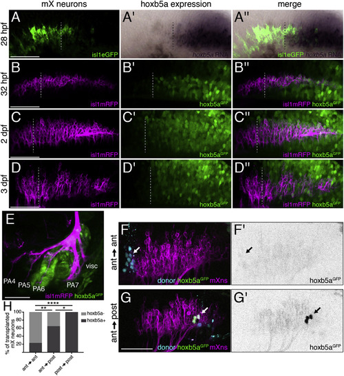
hox5 Expression Distinguishes Anterior and Posterior mX Neurons (A) hoxb5a is expressed in the posterior mX territory. RNA in situ hybridization of 28 hpf embryo for hoxb5a expression (purple) followed by immunostaining against Tg(isl1:eGFPCAAX) to label motor neurons (green) is shown. Dotted lines in (A)–(D) indicate anterior limit of hoxb5a expression. See also Figure S2. (B–E) hoxb5aGFP is expressed in posterior mX neurons, PA6, and PA7. hoxb5aGFP (green) and Tg(isl1:mRFP) (magenta) at 32 hpf (B), 2 dpf (C), or 3 dpf (D and E) are shown. (F and G) Anterior mX neurons marked by lineage dye (cyan) and isl1:mRFP (magenta) transplanted homotopically (F) or heterotopically (G) are hoxb5aGFP-negative (arrow in F) or hoxb5aGFP-positive, respectively (arrow, green in G and black in G′). Host motor neurons express Tg(isl1:mRFP) (magenta). (H) Quantification of (F) and (G). Analysis was done by Fisher’s exact test. Ant → ant: n = 22 neurons, 6 embryos; ant → post: n = 28 neurons, 9 embryos; post → post: n = 13 neurons, 5 embryos. |
| Genes: | |
|---|---|
| Fish: | |
| Anatomical Terms: | |
| Stage Range: | Prim-5 to Protruding-mouth |