
Fig. 6
- ID
- ZDB-FIG-171206-73
- Publication
- Sheets, 2017 - Excessive activation of ionotropic glutamate receptors induces apoptotic hair-cell death independent of afferent and efferent innervation
- Other Figures
- All Figure Page
- Back to All Figure Page
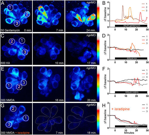
Cytoplasmic Ca2+ dynamics in hair cells treated with gentamycin vs. KA or NMDA. (A,C,E,G) Heat-mapped, time-lapse imaging of ngn1 morphant NMs with hair cells stably expressing GCaMP3. Z-stack images were taken every 30 seconds for 30–50 minutes total. 50 μM gentamycin (A), 600 μM KA (C) or 300 μM NMDA (E,G) was applied 5 minutes into imaging; 10 μM isradipine (G) was applied prior to imaging and remained for the duration. Circles indicate the regions of interest (ROIs) where fluorescence changes in GCaMP3 were measured. Numbers correspond to cells measured for fluorescence intensity traces. Dashed outlines in (H) indicate hair cells; note the reduction in hair-cell number during NMDA exposure. (B,D,F,H) Transformed (ΔF/baseline) fluorescence intensity data. Three cells from each NM are depicted; the red and orange traces correspond to dying hair cells. Cells were chosen to highlight the difference in calcium transients in dying cells exposed to genatmycin (B) vs. iGluR agonists (D,F). Note the reduction in intracellular calcium in (H) does not provide protection from hair-cell death. |
| Gene: | |
|---|---|
| Fish: | |
| Conditions: | |
| Knockdown Reagent: | |
| Anatomical Terms: | |
| Stage: | Day 5 |
| Fish: | |
|---|---|
| Conditions: | |
| Knockdown Reagent: | |
| Observed In: | |
| Stage: | Day 5 |