
FIGURE
Fig. 6
- ID
- ZDB-FIG-171106-4
- Publication
- Radomska et al., 2016 - Characterization and Expression of the Zebrafish qki Paralogs
- Other Figures
- All Figure Page
- Back to All Figure Page
Fig. 6
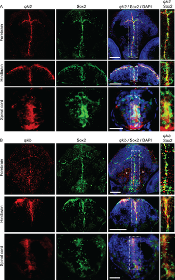
qki2 and qkib are expressed in neural progenitor cells. Representative images of combined in situ hybridization and immunofluorescence detecting qki2 (A) and qkib probes (B), both shown in red. Sox2 antibody is shown in green and DAPI in blue. Images shown are within the forebrain, hindbrain and spinal cord and overlays are denoted in the figure header. Scale bars are 50μm for forebrain and hindbrain images, and 20μm for spinal cord images. Asterisks indicate staining artefacts. |
Expression Data
| Genes: | |
|---|---|
| Fish: | |
| Anatomical Terms: | |
| Stage: | Protruding-mouth |
Expression Detail
Antibody Labeling
Phenotype Data
Phenotype Detail
Acknowledgments
This image is the copyrighted work of the attributed author or publisher, and
ZFIN has permission only to display this image to its users.
Additional permissions should be obtained from the applicable author or publisher of the image.
Full text @ PLoS One