
Fig. 4
- ID
- ZDB-FIG-171018-28
- Publication
- Shim et al., 2017 - Development of zebrafish medulloblastoma-like PNET model by TALEN-mediated somatic gene inactivation
- Other Figures
- All Figure Page
- Back to All Figure Page
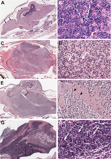
Histopathology of tumors from rb1-TALENs injected tp53 mutant zebrafish. (A) Sagittal section image of H & E staining of wild type zebrafish at 5 month post fertilization. Black arrows and arrow heads indicate forebrain and optic tectum, respectively. Cerebellum and cerebellar granule cells are indicated by white arrows and arrow heads, respectively. (B) High magnitude image of A. Normal cerebellar granule cells have a unique feature that is characterized with small rounded nuclei. (C–H) Sagittal section images of H & E staining of tumors from rb1-TALENs injected tp53 mutant zebrafish. (C) Tumors were mainly arising in cerebellum, medulla, and brainstem. (D) High magnitude image of C. Highly cellular tumor cells with wedged nuclei were observed. Rosette like structures was marked by white arrows. (E) Tumors were mainly arising in cerebellum encompassed a majority of medulla, hypothalamus, and brainstem. The forth ventricle which were surrounded with tumor cells were marked by black arrows. (F) High magnitude image of E. Infiltration of tumor cells was indicated by black arrowheads in dorsal brainstem. (H) High magnitude image of G. Homer-Wright rosettes which are seen in PNETs or medulloblastomas were observed distinctly (white arrows). Anterior is left of all images. Scale bars: 500 μm (A, C, E and G), and 50 μm (B, D, F and H). |
| Fish: | |
|---|---|
| Knockdown Reagent: | |
| Observed In: | |
| Stage: | Adult |