
Fig. 1
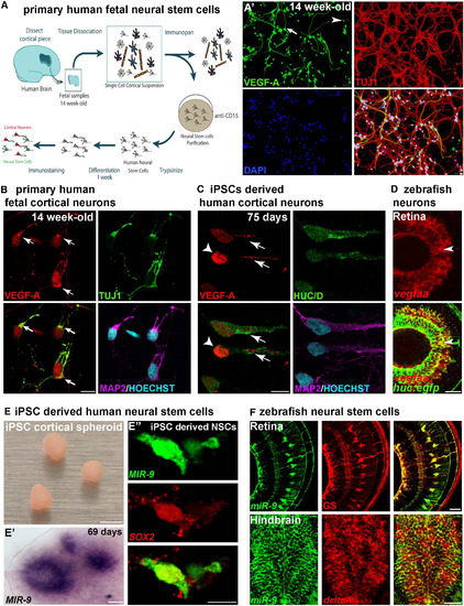
Neuronal VEGF-A and miR-9 Expression in Neural Stem Cells (NSCs) Are Conserved in the Human Brain (A) Schematic representation of the method used to obtain primary human NSCs from fetal brain at 14 weeks of development. At 7 days in culture (A'), a majority of the TUJ1-expressing embryonic neurons express detectable levels of VEGF-A (98.5%, n = 201). NSCs (arrow) and neurons (arrowhead) express VEGF-A in primary human cell culture. (B and C) Confocal sections of primary human cortical neurons (B) and iPSC-derived human cortical neurons (C) immunolabeled with VEGF-A. Young human cortical neurons show heterogeneous localization of VEGF-A (arrows show cytoplasmic and axonal localization; arrowhead indicates strong nuclear expression). (D) Confocal section of double in situ/immunolabeling showing overlap in the expression of vegfaa and Tg(huc:egfp) post-mitotic neurons in the retina (arrowheads). (E) iPSC-derived human cortical spheroid in culture. In situ hybridization showing MIR-9 expression in the ventricular-like zone of human cortical spheroid (E'). Confocal section of double in situ/immunolabeling showing co-localization of MIR-9 and SOX2 in iPSC-derived human NSCs (E''). (F) Confocal section of double immunolabeling with glutamine synthetase (GS) and EGFP in Tg(hsa-MIR-9-2:egfp) retina at 72 hpf, showing miR-9 expression in retinal NSCs. Confocal section of double in situ/immunolabeling showing overlap in the expression of deltaA and EGFP in Tg(hsa-MIR-9-2:egfp) hindbrain at 72 hpf. Dorsal view of the brain with anterior up. Lateral view of the retina. Scale bars: 25 μm in (B) and (C); 100 μm in (D), (E'), and (F); 4 mm in (E); and 10 μm in (A') and (E''). |
| Genes: | |
|---|---|
| Fish: | |
| Anatomical Terms: | |
| Stage Range: | Long-pec to Protruding-mouth |