|
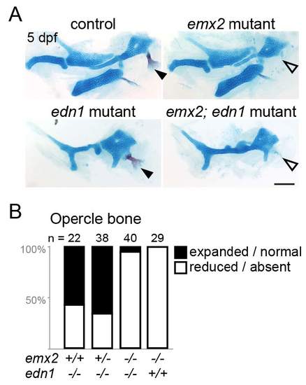
Analysis of emx2; edn1 compound mutants. (A) Alcian blue and Alizarin red staining of control, emx2, edn1, and emx2; edn1 double mutant embryos at 5 dpf. In edn1 mutants, the opercle bone is variably expanded / normal (closed arrowhead) or reduced / absent (open arrowhead). In emx2 and emx2; edn1 mutants, the opercle is almost always reduced or absent. In addition, loss of emx2 fails to rescue the reduced ventral cartilage in edn1 mutants. Scale bar = 100 μm. (B) Quantification of the percentage of opercle bone defects in single and double mutants. The number of sides examined is listed above each bar. The decrease in the proportion of larvae with expanded / normal opercle bone in emx2-/-; edn1-/- mutants compared with emx2+/+; edn1-/- was significant at p < 0.0001 (two-tailed Chi-square).
|