
Fig. 5
- ID
- ZDB-FIG-170501-3
- Publication
- Seberg et al., 2017 - TFAP2 paralogs regulate melanocyte differentiation in parallel with MITF
- Other Figures
- All Figure Page
- Back to All Figure Page
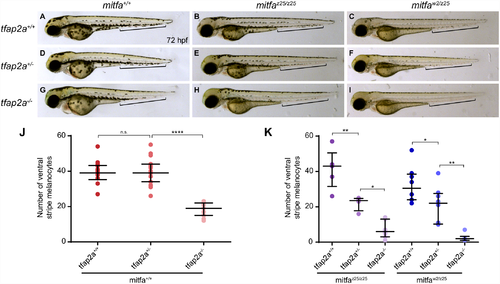
tfap2a and mitfa display a genetic interaction in zebrafish. (A-I) Compared to wildtype embryos at 72 hpf (A), melanocytes in mitfaz25/z25 mutant embryos (B) are dark but punctate, and melanocytes in mitfaw2/z25 trans-heterozygous embryos (C) are pale, punctate, and reduced in number. While heterozygous mutation of tfap2a in the wildtype background (tfap2a+/-) (D) does not result in a melanocyte phenotype, mitfaz25/z25;tfap2a+/- mutants (E) and mitfaw2/z25;tfap2a+/- mutants (F) display a loss of melanocytes in the ventral stripe (brackets). Similarly, the well-characterized phenotype of fewer, paler melanocytes in tfap2a-/- null mutants (G) is more pronounced in both the mitfaz25/z25 mutant background (H) and the mitfaw2/z25 mutant background (I). (J, K) Counts of ventral stripe melanocytes (see brackets in A-I) showed no difference between wildtype and tfap2a+/- embryos (J, n = 10+), while tfap2a+/- embryos in both mitfa mutant backgrounds had significantly fewer melanocytes in the ventral stripe (K, n = 4–8). One-way ANOVA: *p<0.05, **p<0.01, ****p<0.0001. |
| Fish: | |
|---|---|
| Observed In: | |
| Stage: | Protruding-mouth |