
Fig. 1
- ID
- ZDB-FIG-170301-8
- Publication
- Reiten et al., 2017 - Motile-Cilia-Mediated Flow Improves Sensitivity and Temporal Resolution of Olfactory Computations
- Other Figures
- All Figure Page
- Back to All Figure Page
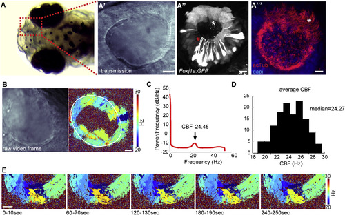
Multiciliated Cells in the Nose Pit of 4-Day-Old Zebrafish Larvae Are Spatially Organized and Beat Their Cilia at a Distinct Frequency (B and C) Fast (100 Hz) transmission microscopy recordings in the nose pit of a representative animal (B, left) showed that the patches of motile cilia beat at frequencies ranging from 20 to 30 Hz (B, right, nose cavity is indicated with a white line), with an average of 24.45 Hz, as shown by power spectral density analysis (C). (D) Average ciliary beating frequency from 130 zebrafish larvae ranges from 19 to 29 Hz, with a median of 24.27 Hz. (E) Beating frequency is stable during 5 min, reported for one representative pit at 1 min intervals. Scale bars represent 10 μm. See also Figures S1 and S2 and Movie S1. |
| Gene: | |
|---|---|
| Antibody: | |
| Fish: | |
| Anatomical Terms: | |
| Stage: | Day 4 |