
FIGURE
Fig. 4
- ID
- ZDB-FIG-161229-8
- Publication
- James et al., 2016 - The Hyaloid Vasculature Facilitates Basement Membrane Breakdown During Choroid Fissure Closure in the Zebrafish Eye
- Other Figures
- All Figure Page
- Back to All Figure Page
Fig. 4
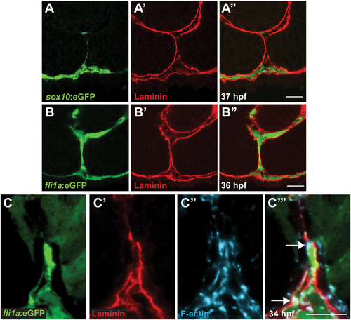
Periocular mesenchymal cells contribute to CFC. (A-C) Sagittal views of the CF stained with anti-GFP (green), Lam-111 (red) and/or phalloidin (blue). (A) Few sox10:eGFP+ cells are detected in the CF, 37 hpf section pictured. (B) fli1a:eGFP+ cells are retained in the CF. 36 hpf section pictured. (C) fli1a:eGFP+ cells possess F-actin accumulations that localize to regions of BM breakdown. Arrows denote puncta of F-actin where Lam-111 is low or absent. 34 hpf section pictured. Scale bar=20 µm (A,B) and 10 µm (C). |
Expression Data
| Antibodies: | |
|---|---|
| Fish: | |
| Anatomical Terms: | |
| Stage Range: | Prim-15 to Prim-25 |
Expression Detail
Antibody Labeling
Phenotype Data
Phenotype Detail
Acknowledgments
This image is the copyrighted work of the attributed author or publisher, and
ZFIN has permission only to display this image to its users.
Additional permissions should be obtained from the applicable author or publisher of the image.
Reprinted from Developmental Biology, 419(2), James, A., Lee, C., Williams, A.M., Angileri, K., Lathrop, K.L., Gross, J.M., The Hyaloid Vasculature Facilitates Basement Membrane Breakdown During Choroid Fissure Closure in the Zebrafish Eye, 262-272, Copyright (2016) with permission from Elsevier. Full text @ Dev. Biol.