
Fig. 3
- ID
- ZDB-FIG-161227-20
- Publication
- Cortes et al., 2015 - Accumulation of the Vitamin D Precursor Cholecalciferol Antagonizes Hedgehog Signaling to Impair Hemogenic Endothelium Formation
- Other Figures
- All Figure Page
- Back to All Figure Page
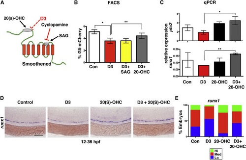
D3 Antagonizes Hh Signaling via Interaction with the Extracellular Domain of Smoothened (A) Diagram depicts the sites of action of Hh pathway modifiers cyclopamine, SAG, and 20-OHC on the Smoothened receptor. The presumptive site of D3 action (red) is shown. (B) FACS of Gli-reporter embryos showed D3 decreased Hh activity (∗p = 0.002). SAG co-treatment (5 μM) did not alter the effect of D3, while 20(S)-OHC (10 μM) partially blocked Hh inhibition (∗∗p = 0.040) (5 embryos/sample × 4 replicates/condition). Error bars, mean ± SD. (C) Co-treatment with 20(S)-OHC corrected D3-mediated reductions in ptc2 (∗p = 0.019) and runx1 (∗∗p = 0.003) by qPCR (40 pooled embryos/condition × 3 replicates). Error bars, mean ± SD. (D) 20(S)-OHC restored runx1 expression in D3-treated embryos. Scale bar, 100 μm. (E) Qualitative phenotypic distribution of embryos from (D) scored for runx1 (n > 20 embryos/condition). |
| Genes: | |
|---|---|
| Fish: | |
| Conditions: | |
| Anatomical Terms: | |
| Stage: | Prim-25 |
| Fish: | |
|---|---|
| Conditions: | |
| Observed In: | |
| Stage: | Prim-25 |