
Fig. 1
- ID
- ZDB-FIG-161227-18
- Publication
- Cortes et al., 2015 - Accumulation of the Vitamin D Precursor Cholecalciferol Antagonizes Hedgehog Signaling to Impair Hemogenic Endothelium Formation
- Other Figures
- All Figure Page
- Back to All Figure Page
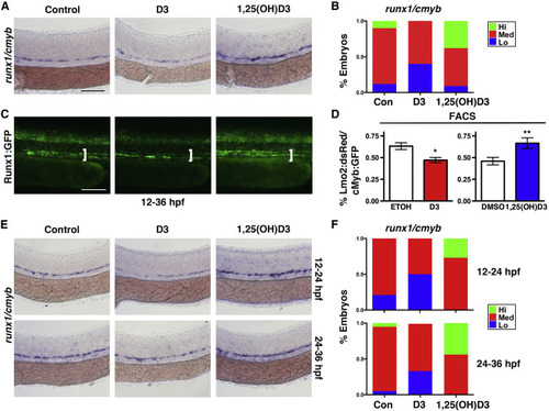
Vitamin D Metabolites Have Differential Effects on HSC Formation (A) Exposure to D3 (50 μM) decreased runx1/cmyb expression by WISH at 36 hpf, and 1,25(OH)D3 (10 μM) increased expression. Scale bar, 100 μm. (B) Qualitative phenotypic distribution of embryos from (A) scored with low, medium, or high runx1/cmyb expression in the AGM (n > 50 embryos/condition). (C) Runx1:GFP embryos exhibited either diminished or enhanced expression with D3 or 1,25(OH)D3 exposure, respectively. Scale bar, 100 μm. (D) FACS analysis of double-positive HSPCs in Lmo2:dsRed/cMyb:GFP embryos confirmed a 25% (∗p = 0.014) decrease with D3 versus a 20% (∗∗p = 0.029) increase with 1,25(OH)D3 compared to controls (5 embryos/sample × 4 replicates/condition). Error bars, mean ± SD. (E) D3 treatment during hemogenic niche specification (12–24 hpf) or HSC induction (24–36 hpf) only decreased runx1/cmyb by WISH in the early exposure window, whereas 1,25(OH)D3 increased expression during either treatment period. Scale as in (A). (F) Qualitative phenotypic distribution of embryos from (E) scored for runx1/cmyb as in (B) (n > 20 embryos/condition). See also Figure S1. |
| Fish: | |
|---|---|
| Conditions: | |
| Observed In: | |
| Stage: | Prim-25 |