
FIGURE
Fig. 1
- ID
- ZDB-FIG-160927-19
- Publication
- Jim et al., 2016 - Infection of zebrafish embryos with live fluorescent Streptococcus pneumoniae as a real-time pneumococcal meningitis model
- Other Figures
- All Figure Page
- Back to All Figure Page
Fig. 1
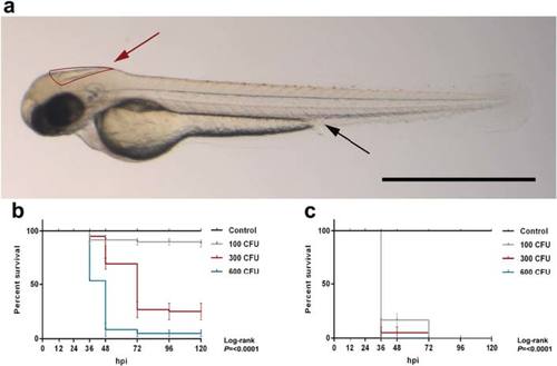
Survival curves of 2 days post-fertilization embryos injected through different routes with wild-type Streptococcus pneumoniae D39. a Casper zebrafish embryo at 2 days post - fertilization. Red arrow indicates the hindbrain ventricle infection route, and black arrow indicates the caudal vein infection route. Scale bar, 500 µm. b, c Injection in the b hindbrain ventricle (HBV) or c caudal vein with indicated doses. Hpi hours post injection, CFU colony-forming units. The data represent the mean ± SEM of three individual experiments with 20 embryos in each group |
Expression Data
Expression Detail
Antibody Labeling
Phenotype Data
| Fish: | |
|---|---|
| Condition: | |
| Observed In: | |
| Stage Range: | Long-pec to Day 6 |
Phenotype Detail
Acknowledgments
This image is the copyrighted work of the attributed author or publisher, and
ZFIN has permission only to display this image to its users.
Additional permissions should be obtained from the applicable author or publisher of the image.
Full text @ J Neuroinflammation