
Fig. 1
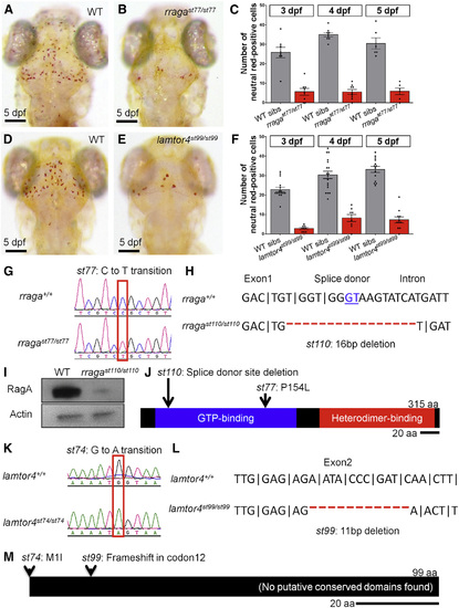
Mutational Analysis Demonstrates that rraga and lamtor4 Are Essential for Microglia Development (A–F) Comparison of microglia numbers in rragast77/st77 mutants, lamtor4st99/st99 mutants, and WT siblings. (A, B, D, and E) Neutral red-stained larvae of the indicated genotypes at 5 dpf are shown. Dorsal views, scale bar, 50 µm. (C and F) Quantifications of neutral red-stained microglia in rraga (C) and lamtor4 (F) mutants at 3, 4, and 5 dpf are shown. (G–J) Molecular analysis of mutations in rraga. (G) Sequence chromatograms show point mutation in rragast77 mutation. (H) Sequence deleted in rragast110 mutation shows exon-intron boundary and deleted splice donor. (I) Immunoblot showing pronounced reduction of RagA protein in rragast77/st77 mutants at 5 dpf. Actin is shown as a loading control. (J) Schematic of RagA protein shows conserved domains and positions of the mutant lesions. (K–M) Molecular analysis of mutations in lamtor4. (K) Sequence chromatograms show point mutation in lamtor4st74 mutation, which changes the ATG initiation codon to ATA. (L) Sequence deleted in lamtor4st99 mutation shows the reading frame in WT and the alteration caused by the 11-bp deletion in the mutation. (M) Schematic of the Lamtor4 protein shows positions of the mutant lesions. Larvae shown in (A), (B), (D), and (E) were genotyped by PCR after photography. |
| Gene: | |
|---|---|
| Antibodies: | |
| Fish: | |
| Condition: | |
| Anatomical Term: | |
| Stage: | Day 5 |
| Fish: | |
|---|---|
| Condition: | |
| Observed In: | |
| Stage Range: | Protruding-mouth to Day 5 |