
FIGURE
Fig. S3
- ID
- ZDB-FIG-160112-2
- Publication
- Zang et al., 2015 - Recoverin depletion accelerates cone photoresponse recovery
- Other Figures
- All Figure Page
- Back to All Figure Page
Fig. S3
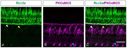
Co-staining of Rcv2a and PKC Antibodies on Retina Sections. Z-projections of confocal image stacks of immunochemical staining on adult retinas. White arrowhead marked the bipolar cell which was labeled by both Rcv2a and PKC antibodies. Yellow arrowhead marked the bipolar cell which was only labeled by Rcv2a antibody. Blue arrowhead marked the ON-bipolar cell which was only labeled by PKC antibody. Scale bar=20 µm. |
Expression Data
| Antibodies: | |
|---|---|
| Fish: | |
| Anatomical Terms: | |
| Stage: | Adult |
Expression Detail
Antibody Labeling
Phenotype Data
Phenotype Detail
Acknowledgments
This image is the copyrighted work of the attributed author or publisher, and
ZFIN has permission only to display this image to its users.
Additional permissions should be obtained from the applicable author or publisher of the image.
Full text @ Open Biol.