|
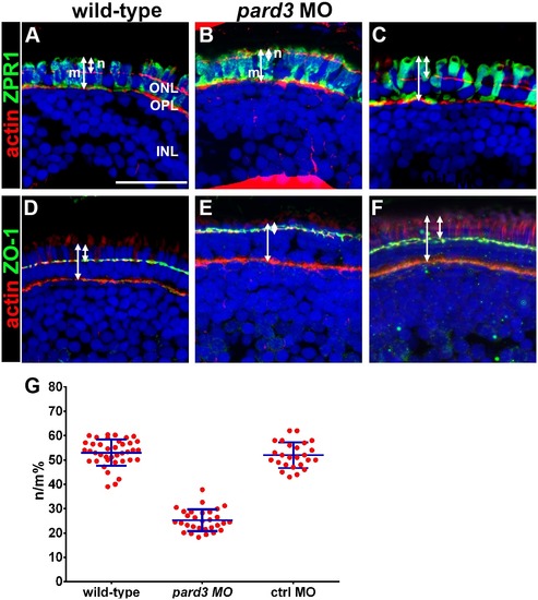
Apical domain size is reduced in pard3 morphants. (A–C) Transverse cryosections of 5 dpf retinas were stained with phalloidin to label actin (red) and zpr-1 to label double cones (green). The length of the photoreceptor (m) and the length of the apical region of the inner segment (n) are noted by arrows. (D–F) Transverse sections stained with phalloidin (red) and ZO-1 (green) to label the outer limiting membrane. Sections were also counterstained with DAPI (blue). (G) Quantification of apical domain as a percentage of total photoreceptor inner segment length. All values are plotted as a percentage of n/m, as defined in the text. Wild type = 53%; pard3 morphants = 25%; control morphants = 52%. Blue error bars indicate the mean ± standard deviation. ONL = outer nuclear layer; OPL = outer plexiform layer; INL = inner nuclear layer. Scale bar = 20 μm.
|