
Fig. 1
- ID
- ZDB-FIG-150612-8
- Publication
- Nussbaum et al., 2013 - Homeostatic generation of reactive oxygen species protects the zebrafish liver from steatosis
- Other Figures
- All Figure Page
- Back to All Figure Page
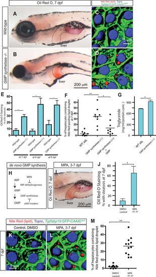
Lack of de novo GMP synthesis leads to hepatic steatosis. (A,B) Lateral views of wild-type sibling and GMP synthetases850 mutant larvae stained with Oil Red O (ORO) at 7 dpf. GMP synthetases850 mutant larvae develop liver steatosis at 7 dpf. Broken lines outline the liver. (C,D) Projected confocal images of the wild-type sibling (C) and GMP synthetases850 mutant (D) liver visualized for Nile Red (red) and To-pro-3 (blue) staining and Tg (fabp10:GFP-CAAX)lri1 (green) expression at 7 dpf. In GMP synthetases850 mutant larvae, lipid droplets stained by Nile Red are evident in hepatocytes. (E) The percentage of wild-type and GMP synthetases850 mutant larvae showing liver steatosis scored by whole-mount ORO staining at 7, 8, and 9 dpf. ORO staining experiments with wild-type larvae at 7, 8, and 9 dpf were repeated five times (total n = 138 larvae examined and total n = 4 showed ORO signal in the liver), five times (total n = 74 larvae examined and total n = 9 showed ORO signal in the liver), and six times (total n = 67 larvae examined and total n = 13 showed ORO signal in the liver), respectively. ORO staining experiments with GMP synthetases850 mutant larvae at 7, 8, and 9 dpf were repeated five times (total n = 124 larvae examined and total n = 36 showed ORO signal in the liver), four times (total n = 34 larvae examined and total n = 15 showed ORO signal in the liver), and three times (total n = 26 larvae examined and total n = 12 showed ORO signal in the liver), respectively. (F) Quantification of liver steatosis measured by the percentage of hepatocytes containing Nile Red positive lipid droplets in wild-type sibling (n = 9), GMP synthetases850 mutant (n = 9), and 150 µM GMP-treated GMP synthetases850 mutant larvae (n = 9). The percentage of hepatocytes containing lipid droplets is significantly increased in GMP synthetases850 mutant larvae. (G) Triglyceride levels measured in whole-body extracts of wild-type and GMP synthetases850 mutant larvae at 7 dpf. (H) A schematic of the de novo GMP synthesis pathway. In the linear de novo GMP synthesis pathway, IMP is converted to XMP by IMP dehydrogenase, and subsequently, XMP is converted to GMP by GMP synthetase. MPA inhibits the function of IMP dehydrogenase. The s850 mutation disrupts the function of GMP synthetase. (I) Wild-type larvae exposed to MPA from 3 to 7 dpf were fixed and stained with ORO at 7 dpf. MPA-treated larvae developed liver steatosis. (J) The percentage of control DMSO or MPA-treated larvae showing liver steatosis scored by whole-mount ORO staining at 7 dpf. MPA treatment increased the percentage of larvae showing liver steatosis. ORO staining experiments with MPA-treated larvae were repeated three times with an average n = 35.4 larvae per experiment (total n = 106 larvae examined and total n = 70 larvae showed ORO signal in the liver). (K,L) Projected confocal images of wild-type larvae treated with DMSO (K) or MPA (L) from 3 to 7 dpf visualized for Nile Red (red) and To-pro-3 (blue) staining and Tg (fabp10:GFP-CAAX)lri1 expression (green). Nile Red stains lipid droplets, and Topro stains nuclei. (M) Quantification of liver steatosis measured by the percentage of hepatocytes containing lipid droplets in DMSO-treated control and MPA-treated larvae at 7 dpf. MPA-treatment significantly increased the percentage of hepatocytes containing lipid droplets. sb, swim bladder. *P < 0.05, **P < 0.01; error bars indicate SD. |
| Gene: | |
|---|---|
| Fish: | |
| Condition: | |
| Anatomical Term: | |
| Stage: | Days 7-13 |