
Fig. 4
- ID
- ZDB-FIG-150330-33
- Publication
- Fukui et al., 2014 - S1P-Yap1 Signaling Regulates Endoderm Formation Required for Cardiac Precursor Cell Migration in Zebrafish
- Other Figures
- All Figure Page
- Back to All Figure Page
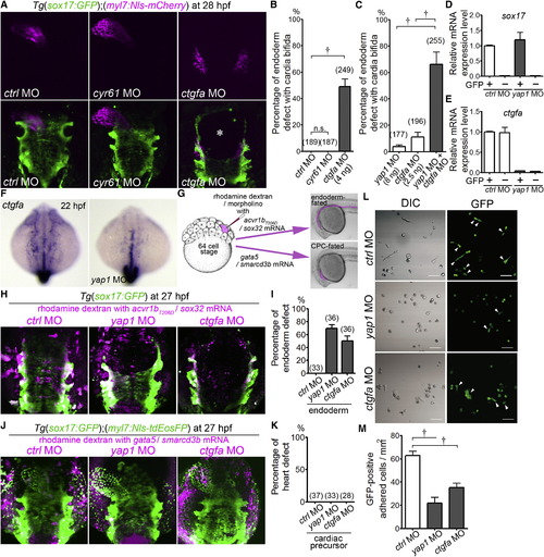
Endodermal Cells Cell-Autonomously Regulate Their Survival in a Manner Dependent on the Expression of Yap1-Promoted ctgfa (A) 3D-rendered two-photon laser-scanned z-stack images of Tg(sox17:GFP);(myl7:Nls-mCherry) embryos (28 hpf) injected with MO indicated at the bottom. An asterisk shows the defect of the endoderm. Dorsal view. (B and C) Incidence of endoderm defects with cardia bifida of Tg(sox17:GFP);(myl7:Nls-mCherry) embryos injected with MO indicated at the bottom. Total number of the embryos analyzed in each morphant group is indicated at the top of the column. (D and E) Quantitative-PCR analyses of expression of sox17 mRNA (D) and ctgfa mRNA (E) in the endoderm (GFP+ cells) and extra-endoderm (GFP- cells) of the Tg(sox17:GFP) embryos injected with MO indicated at the bottom. GFP+ and GFP- cells from the embryo at 12 hpf were sorted by GFP fluorescence (n = 4). (F) WISH analyses of ctgfa mRNA expression in the wild-type embryo (left) and in the yap1 morphant (right; 22 hpf). A set of representative images of four independent experiments is shown. Dorsal view. (G) Schematic illustration of injection of MO into the endoderm-fated cells (simultaneous injection of both acvr1bT206D and sox32 mRNAs) and into the CPC-fated cells (simultaneous injection of both gata5 and smarcd3b mRNAs) with rhodamine dextran to visualize the cells injected with MO and mRNAs. MO, mRNA, and rhodamine dextran were coinjected into one blastomere at the 64-cell stage. (H) Representative 3D-rendered confocal images of the Tg(sox17:GFP) embryo injected with mRNAs indicated at the top with the MOs indicated above the image as explained in (G). (I) Quantitative analyses of incidence of endodermal defects in the embryos examined in (H). (J) Representative 3D-rendered confocal images of the Tg(sox17:GFP);(myl7:Nls-tdEosFP) embryo injected with mRNAs indicated at the top with the MOs indicated above the image as explained in (G). (K) Quantitative analyses of incidence of heart defects in the embryos examined in (J). (L and M) Attachment and spread of the endodermal cells obtained at 12 hpf from Tg(sox17:GFP) embryos injected with MO indicated at the top to the fibronectin-coated dish. Arrowheads indicate the cells spreading after attachment to the dish. A panel of representative result of three independent experiments is shown in (L). Scale bars represent 50 µm. (M) The graph shows the number of the spreading GFP-positive cells after attachment. †p < 0.01. See also Figure S4. |
| Genes: | |
|---|---|
| Fish: | |
| Knockdown Reagents: | |
| Anatomical Terms: | |
| Stage Range: | 5-9 somites to Prim-5 |
| Fish: | |
|---|---|
| Knockdown Reagents: | |
| Observed In: | |
| Stage Range: | 5-9 somites to Prim-5 |
Reprinted from Developmental Cell, 31, Fukui, H., Terai, K., Nakajima, H., Chiba, A., Fukuhara, S., Mochizuki, N., S1P-Yap1 Signaling Regulates Endoderm Formation Required for Cardiac Precursor Cell Migration in Zebrafish, 128-136, Copyright (2014) with permission from Elsevier. Full text @ Dev. Cell