
Fig. 4
- ID
- ZDB-FIG-150121-16
- Publication
- Wanglar et al., 2014 - Tbx Protein Level Critical for Clock-Mediated Somite Positioning Is Regulated through Interaction between Tbx and Ripply
- Other Figures
- All Figure Page
- Back to All Figure Page
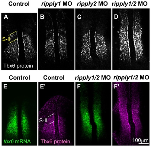
Proper positioning of Tbx6 domain depends on ripply. (A-D) Patterns of Tbx6 protein at the 8 somite stage in control (n = 20) (A), ripply1 morphant (n = 25) (B), ripply2 morphant (n = 20) (C) and ripply1/ripply2 double morphant (n = 30) (D). Comparison of Tbx6 protein (E′, F′) with its mRNA (E, F) patterns in control (E, E′) and ripply1/ripply2 double morphant (F, F′). ripply1 morphants show graded expansion of Tbx6 protein anteriorly (B) but ripply2 morphants (C) show no significant difference from control embryos (A). Double knockdown of ripply1 and ripply2 show strong expansion of Tbx6 protein anteriorly (D). In the double morphants, tbx6 mRNA is also anteriorly expanded to some level, but not so significantly as Tbx6 protein (F, F′). A total of 20 injected embryos were observed for each injection. While ripply2 morphant appeared similar to control embryos in Tbx6 protein pattern, 100% of the ripply1 morphants and 100% of the ripply1 and ripply2 double morphants displayed anterior expansion of Tbx6 protein shown in (B) and (D) respectively. The dotted lines indicate S-II (A, E′) region. |
| Gene: | |
|---|---|
| Antibody: | |
| Fish: | |
| Knockdown Reagents: | |
| Anatomical Terms: | |
| Stage: | 5-9 somites |