
FIGURE
Fig. 7
Fig. 7
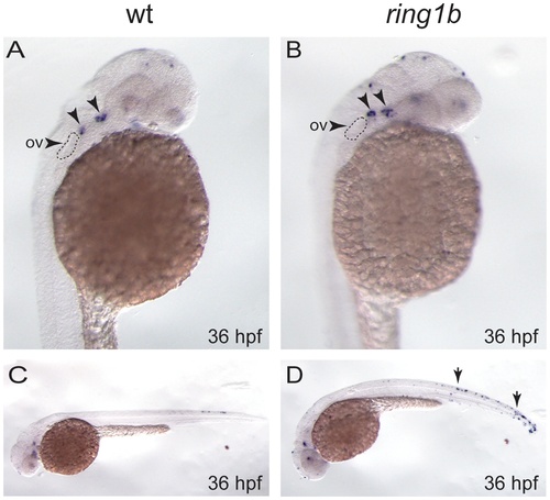
Apoptosis is slightly increased in the pharyngeal arch of ring1b mutants. Lateral views of WT and ring1b embryos at 36 hpf stained for TUNEL. In WT embryos two small clusters of TUNEL-positive apoptotic cells were detected in the pharyngeal arch region just posterior to the eye (A, arrows). These clusters appear to contain more apoptotic cells in the ring1b mutants (B, arrows). The arrowhead indicates the otic vesicle (ov). WT embryos at 36 hpf contain few apoptotic cells in the trunk (C), whereas there is an increase in overall apoptosis particularly in the trunk and the tail in ring1b mutants (D, arrows). |
Expression Data
Expression Detail
Antibody Labeling
Phenotype Data
| Fish: | |
|---|---|
| Observed In: | |
| Stage: | Prim-25 |
Phenotype Detail
Acknowledgments
This image is the copyrighted work of the attributed author or publisher, and
ZFIN has permission only to display this image to its users.
Additional permissions should be obtained from the applicable author or publisher of the image.
Full text @ PLoS One