
Fig. 1
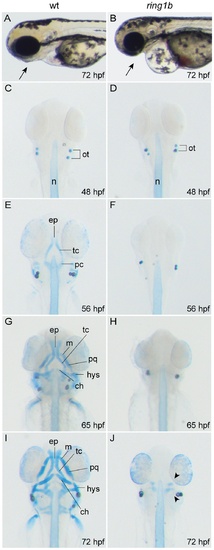
Ring1b mutants lack almost all head cartilage elements. Lateral view of WT and ring1b live embryos at 72 hpf (A, B). Alcian-Blue stained head cartilages of WT (C, E, G and H) and ring1b (D, F, H and J) mutants at the indicated developmental points, ventral views. The paired trabeculae have elongated and fused posteriorly in WT embryos at 56 hpf (E) and by 72 hpf the elaborate cartilagenous skeleton of the head has been established (I). In contrast, no cartilage is visible in ring1b mutants except for two minute cartilage deposits at 72 hpf ring1b mutants (J: arrowheads). ch: ceratohyal; ep: ethmoid plate; hys: hyosymplectic; m: Meckel’s cartilage; pc: parachordal, pq: palatoquadrate; tc: trabeculae. |
| Fish: | |
|---|---|
| Observed In: | |
| Stage Range: | Long-pec to Protruding-mouth |