|
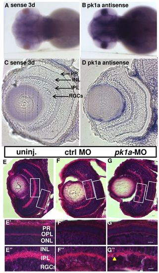
pk1a is expressed in the brain and retina and is not required for overall retinal lamination. (A,B) Whole-mount in situ hybridization of 3 dpf embryos. Images are from the dorsal view. An antisense RNA riboprobe is used to determine the expression pattern of pk1a, and a riboprobe from the sense strand is used as a control. (C,D) Sections of the eye after whole-mount in situ hybridization at 3 dpf: sense control (C) and pk1a antisense (D). Arrows show different layers in the retina. (E–G′′) H&E staining of eye sections at 76–78 hpf. (E–G) Whole retina sections of uninjected (E), control-MO-injected (F) and pk1a-MO-injected larvae (G); magnification 40×. (E′–G′′) Enlarged areas of the white boxes in E–G; the arrowhead in G′′ indicates nuclei disorganization; magnification 63×. PR, photo-receptors; INL, inner nuclear layer; IPL, inner plexiform layer; RGCs, retina ganglion cells; OPL, outer plexiform layer; ONL, outer nuclear layer. Scale bars: 5 μm.
|